Dirican Abuzer, Unal Bulent, Kayaalp Cuneyt, Kirimlioglu Vedat
Department of General Surgery, Medical Faculty of Inonu University, Malatya, Turkey.
J Med Case Rep. 2008 Aug 13;2:273. doi: 10.1186/1752-1947-2-273.
Hydatid cyst disease is common in some regions of the world and is usually located in the liver and lungs. This report presents two cases of primary hydatid cysts located subcutaneously: one in the medial thigh and one in the left palm between the index and middle fingers.
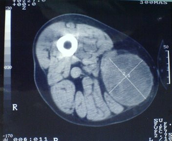
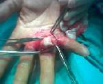
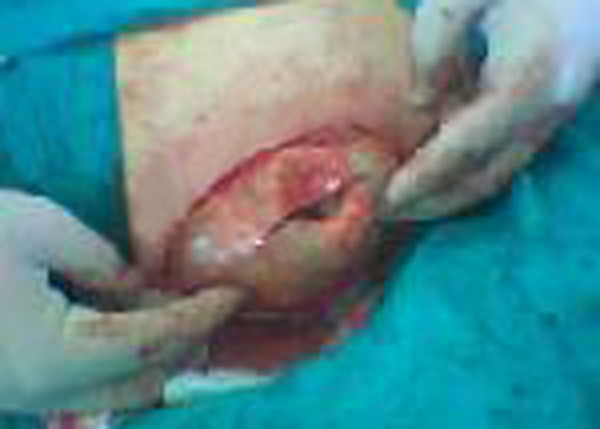
A 64-year-old male farmer visited our hospital because a swelling on the right medial thigh had grown during the last year. Superficial ultrasound and computed tomography revealed a lesion resembling a hydatid cyst. A germinative membrane was encountered during surgical excision. Pathological examination was compatible with a hydatid cyst. The second case involved a 67-year-old male farmer who complained of a swelling that had grown in his left palm in the last year. The preliminary diagnosis was a lipoma. However, a hydatid cyst was diagnosed during surgical excision and after the pathological examination. The patient did not have a history of hydatid cyst disease and hydatid cysts were not detected in other organs. There has been no disease recurrence after following both patients for 3 years.
A hydatid cyst should be considered in the differential diagnosis of subcutaneous cystic lesions in regions where hydatid cysts are endemic, and should be excised totally, with an intact wall, to avoid recurrence.
包虫囊肿病在世界某些地区较为常见,通常位于肝脏和肺部。本报告介绍了两例原发性皮下包虫囊肿病例:一例位于大腿内侧,另一例位于左手食指和中指之间的手掌处。
一名64岁男性农民因右大腿内侧肿物在过去一年中逐渐增大前来我院就诊。浅表超声和计算机断层扫描显示一个类似包虫囊肿的病变。手术切除过程中发现了生发层膜。病理检查结果与包虫囊肿相符。第二例患者为一名67岁男性农民,他主诉左手掌肿物在过去一年中逐渐增大。初步诊断为脂肪瘤。然而,手术切除及病理检查后诊断为包虫囊肿。该患者既往无包虫囊肿病史,其他器官未检测到包虫囊肿。对两名患者随访3年,均未出现疾病复发。
在包虫囊肿流行地区,皮下囊性病变的鉴别诊断中应考虑包虫囊肿,应完整切除囊肿壁以避免复发。