• 文献检索
  • 文档翻译
  • 深度研究
  • 学术资讯
  • Suppr Zotero 插件Zotero 插件
  • 邀请有礼
  • 套餐&价格
  • 历史记录
应用&插件
Suppr Zotero 插件Zotero 插件浏览器插件Mac 客户端Windows 客户端微信小程序
定价
高级版会员购买积分包购买API积分包
服务
文献检索文档翻译深度研究API 文档MCP 服务
关于我们
关于 Suppr公司介绍联系我们用户协议隐私条款
关注我们

Suppr 超能文献

核心技术专利:CN118964589B侵权必究
粤ICP备2023148730 号-1Suppr @ 2026

文献检索

告别复杂PubMed语法,用中文像聊天一样搜索,搜遍4000万医学文献。AI智能推荐,让科研检索更轻松。

立即免费搜索

文件翻译

保留排版,准确专业,支持PDF/Word/PPT等文件格式,支持 12+语言互译。

免费翻译文档

深度研究

AI帮你快速写综述,25分钟生成高质量综述,智能提取关键信息,辅助科研写作。

立即免费体验

《小鼠肢体解剖图谱》:用于研究胚胎肢体模式形成的交互式3D工具。

The Mouse Limb Anatomy Atlas: an interactive 3D tool for studying embryonic limb patterning.

作者信息

Delaurier April, Burton Nicholas, Bennett Michael, Baldock Richard, Davidson Duncan, Mohun Timothy J, Logan Malcolm Po

机构信息

Division of Developmental Biology, National Institute for Medical Research, The Ridgeway, Mill Hill, London, UK.

出版信息

BMC Dev Biol. 2008 Sep 15;8:83. doi: 10.1186/1471-213X-8-83.

DOI:10.1186/1471-213X-8-83
PMID:18793391
原文链接:https://pmc.ncbi.nlm.nih.gov/articles/PMC2553786/
Abstract

BACKGROUND

The developing mouse limb is widely used as a model system for studying tissue patterning. Despite this, few references are available that can be used for the correct identification of developing limb structures, such as muscles and tendons. Existing textual references consist of two-dimensional (2D) illustrations of the adult rat or mouse limb that can be difficult to apply when attempting to describe the complex three-dimensional (3D) relationship between tissues.

RESULTS

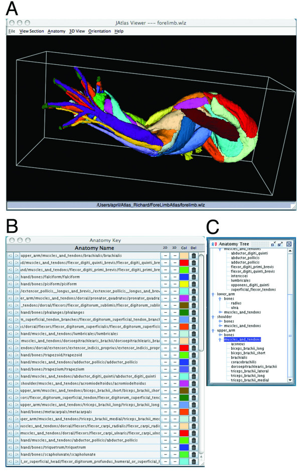
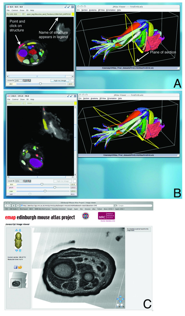
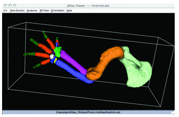
To improve the resources available in the mouse model, we have generated a free, web-based, interactive reference of limb muscle, tendon, and skeletal structures at embryonic day (E) 14.5 http://www.nimr.mrc.ac.uk/3dlimb/. The Atlas was generated using mouse forelimb and hindlimb specimens stained using immunohistochemistry to detect muscle and tendon. Limbs were scanned using Optical Projection Tomography (OPT), reconstructed to make 3D models and annotated using computer-assisted segmentation tools in Amira 3D Visualisation software. The annotated dataset is visualised using Java, JAtlasView software. Users click on the names of structures and view their shape, position and relationship with other structures within the 3D model and also in 2D virtual sections.

CONCLUSION

The Mouse Limb Anatomy Atlas provides a novel and valuable tool for researchers studying limb development and can be applied to a range of research areas, including the identification of abnormal limb patterning in transgenic lines and studies of models of congenital limb abnormalities. By using the Atlas for "virtual" dissection, this resource offers an alternative to animal dissection. The techniques we have developed and employed are also applicable to many other model systems and anatomical structures.

摘要

背景

发育中的小鼠肢体被广泛用作研究组织模式形成的模型系统。尽管如此,可用于正确识别发育中的肢体结构(如肌肉和肌腱)的参考文献却很少。现有的文字参考文献包括成年大鼠或小鼠肢体的二维(2D)插图,在试图描述组织之间复杂的三维(3D)关系时可能难以应用。

结果

为了改进小鼠模型中的可用资源,我们生成了一个免费的、基于网络的、关于胚胎第14.5天(E14.5)肢体肌肉、肌腱和骨骼结构的交互式参考文献http://www.nimr.mrc.ac.uk/3dlimb/。该图谱是使用免疫组织化学染色以检测肌肉和肌腱的小鼠前肢和后肢标本生成的。使用光学投影断层扫描(OPT)对肢体进行扫描,重建以制作3D模型,并在Amira 3D可视化软件中使用计算机辅助分割工具进行注释。注释后的数据集使用Java、JAtlasView软件进行可视化。用户点击结构名称,可在3D模型以及2D虚拟切片中查看其形状、位置以及与其他结构的关系。

结论

小鼠肢体解剖图谱为研究肢体发育的研究人员提供了一种新颖且有价值的工具,可应用于一系列研究领域,包括鉴定转基因品系中的异常肢体模式以及先天性肢体异常模型的研究。通过使用该图谱进行“虚拟”解剖,此资源提供了一种替代动物解剖的方法。我们开发和采用的技术也适用于许多其他模型系统和解剖结构。

https://cdn.ncbi.nlm.nih.gov/pmc/blobs/c802/2553786/b668f82c1fa9/1471-213X-8-83-3.jpg
https://cdn.ncbi.nlm.nih.gov/pmc/blobs/c802/2553786/62c96b1a06e4/1471-213X-8-83-1.jpg
https://cdn.ncbi.nlm.nih.gov/pmc/blobs/c802/2553786/c9044231e24b/1471-213X-8-83-2.jpg
https://cdn.ncbi.nlm.nih.gov/pmc/blobs/c802/2553786/b668f82c1fa9/1471-213X-8-83-3.jpg
https://cdn.ncbi.nlm.nih.gov/pmc/blobs/c802/2553786/62c96b1a06e4/1471-213X-8-83-1.jpg
https://cdn.ncbi.nlm.nih.gov/pmc/blobs/c802/2553786/c9044231e24b/1471-213X-8-83-2.jpg
https://cdn.ncbi.nlm.nih.gov/pmc/blobs/c802/2553786/b668f82c1fa9/1471-213X-8-83-3.jpg

相似文献

1
The Mouse Limb Anatomy Atlas: an interactive 3D tool for studying embryonic limb patterning.《小鼠肢体解剖图谱》:用于研究胚胎肢体模式形成的交互式3D工具。
BMC Dev Biol. 2008 Sep 15;8:83. doi: 10.1186/1471-213X-8-83.
2
Pitx1 determines the morphology of muscle, tendon, and bones of the hindlimb.Pitx1决定后肢肌肉、肌腱和骨骼的形态。
Dev Biol. 2006 Nov 1;299(1):22-34. doi: 10.1016/j.ydbio.2006.06.055. Epub 2006 Jul 14.
3
Tbx4 and tbx5 acting in connective tissue are required for limb muscle and tendon patterning.Tbx4 和 tbx5 在结缔组织中的作用对于肢体肌肉和肌腱的模式形成是必需的。
Dev Cell. 2010 Jan 19;18(1):148-56. doi: 10.1016/j.devcel.2009.11.013.
4
Tendons and muscles of the mouse forelimb during embryonic development.小鼠胚胎发育期间前肢的肌腱和肌肉
Dev Dyn. 2009 Mar;238(3):693-700. doi: 10.1002/dvdy.21866.
5
Manifestation of the limb prepattern: limb development in the absence of sonic hedgehog function.肢体预模式的表现:在缺乏音猬因子功能的情况下的肢体发育。
Dev Biol. 2001 Aug 15;236(2):421-35. doi: 10.1006/dbio.2001.0346.
6
Recent molecular advances in understanding vertebrate limb development.脊椎动物肢体发育研究的近期分子进展。
Br J Plast Surg. 1997 Feb;50(2):109-15. doi: 10.1016/s0007-1226(97)91322-9.
7
Vertebrate limb development and possible clues to diversity in limb form.脊椎动物肢体发育及肢体形态多样性的可能线索。
J Morphol. 2002 Apr;252(1):29-37. doi: 10.1002/jmor.10016.
8
Tissue patterning in the developing mouse limb.发育中小鼠肢体的组织模式形成
Int J Dev Biol. 1990 Sep;34(3):323-36.
9
JAtlasView: a Java atlas-viewer for browsing biomedical 3D images and atlases.JAtlasView:一款用于浏览生物医学3D图像和图谱的Java图谱查看器。
BMC Bioinformatics. 2005 Mar 9;6:47. doi: 10.1186/1471-2105-6-47.
10
Tbx5 is essential for forelimb bud initiation following patterning of the limb field in the mouse embryo.在小鼠胚胎中,肢体区域形成模式后,Tbx5对于前肢芽的起始至关重要。
Development. 2003 Feb;130(3):623-33. doi: 10.1242/dev.00191.

引用本文的文献

1
Harmonizing mouse anatomy terminology: a common language?统一小鼠解剖学术语:一种通用语言?
Mamm Genome. 2025 Sep 10. doi: 10.1007/s00335-025-10156-6.
2
A novel biomechanical model of the proximal mouse forelimb predicts muscle activity in optimal control simulations of reaching movements.一种新型的小鼠前肢近端生物力学模型可预测伸手运动最优控制模拟中的肌肉活动。
J Neurophysiol. 2025 Apr 1;133(4):1266-1278. doi: 10.1152/jn.00499.2024. Epub 2025 Mar 18.
3
FARMS: Framework for Animal and Robot Modeling and Simulation.FARMS:动物与机器人建模及仿真框架

本文引用的文献

1
FishNet: an online database of zebrafish anatomy.FishNet:斑马鱼解剖学在线数据库。
BMC Biol. 2007 Aug 17;5:34. doi: 10.1186/1741-7007-5-34.
2
Generation of transgenic tendon reporters, ScxGFP and ScxAP, using regulatory elements of the scleraxis gene.利用硬骨素基因的调控元件生成转基因肌腱报告基因ScxGFP和ScxAP。
Dev Dyn. 2007 Jun;236(6):1677-82. doi: 10.1002/dvdy.21179.
3
Ultramicroscopy: three-dimensional visualization of neuronal networks in the whole mouse brain.超微显微镜:全脑小鼠神经网络的三维可视化
bioRxiv. 2025 Feb 21:2023.09.25.559130. doi: 10.1101/2023.09.25.559130.
4
Three-dimensional visualization and quantitative analysis of embryonic and fetal thigh muscles using magnetic resonance and phase-contrast X-ray imaging.利用磁共振和相位对比 X 射线成像对胚胎和胎儿大腿肌肉进行三维可视化和定量分析。
J Anat. 2022 Dec;241(6):1310-1323. doi: 10.1111/joa.13764. Epub 2022 Sep 19.
5
A Whole-Body Musculoskeletal Model of the Mouse.小鼠的全身肌肉骨骼模型。
IEEE Access. 2021;9:163861-163881. doi: 10.1109/access.2021.3133078. Epub 2021 Dec 6.
6
A Tendon-Specific Double Reporter Transgenic Mouse Enables Tracking Cell Lineage and Functions Alteration In Vitro and In Vivo.一种肌腱特异性双报告基因转基因小鼠能够在体外和体内追踪细胞谱系及功能改变。
Int J Mol Sci. 2021 Oct 17;22(20):11189. doi: 10.3390/ijms222011189.
7
Modulation of tactile feedback for the execution of dexterous movement.灵巧运动执行中的触觉反馈调节。
Science. 2021 Oct 15;374(6565):316-323. doi: 10.1126/science.abh1123. Epub 2021 Oct 14.
8
Is Essential for Patterning of Multiple Musculoskeletal Tissues but Dispensable for Tendon Differentiation.对于多种肌肉骨骼组织的模式形成是必需的,但对于肌腱分化是可有可无的。
Stem Cells Dev. 2021 Jun 1;30(11):601-609. doi: 10.1089/scd.2020.0209. Epub 2021 Apr 27.
9
4D formation of human embryonic forelimb musculature.人类胚胎前肢肌肉的 4D 形成。
Development. 2021 Feb 17;148(4):dev194746. doi: 10.1242/dev.194746.
10
Individual Limb Muscle Bundles Are Formed through Progressive Steps Orchestrated by Adjacent Connective Tissue Cells during Primary Myogenesis.个体肢体肌束通过初级肌发生过程中相邻结缔组织细胞的有序渐进步骤形成。
Cell Rep. 2020 Mar 10;30(10):3552-3565.e6. doi: 10.1016/j.celrep.2020.02.037.
Nat Methods. 2007 Apr;4(4):331-6. doi: 10.1038/nmeth1036. Epub 2007 Mar 25.
4
Pitx1 determines the morphology of muscle, tendon, and bones of the hindlimb.Pitx1决定后肢肌肉、肌腱和骨骼的形态。
Dev Biol. 2006 Nov 1;299(1):22-34. doi: 10.1016/j.ydbio.2006.06.055. Epub 2006 Jul 14.
5
Four-dimensional ultrasonography of the fetal heart using a novel Tomographic Ultrasound Imaging display.使用新型断层超声成像显示技术对胎儿心脏进行四维超声检查。
J Perinat Med. 2006;34(1):39-55. doi: 10.1515/JPM.2006.006.
6
Case study: an evaluation of user-assisted hierarchical watershed segmentation.案例研究:用户辅助的分层分水岭分割评估
Med Image Anal. 2005 Dec;9(6):566-78. doi: 10.1016/j.media.2005.04.007.
7
JAtlasView: a Java atlas-viewer for browsing biomedical 3D images and atlases.JAtlasView:一款用于浏览生物医学3D图像和图谱的Java图谱查看器。
BMC Bioinformatics. 2005 Mar 9;6:47. doi: 10.1186/1471-2105-6-47.
8
Rapid high resolution three dimensional reconstruction of embryos with episcopic fluorescence image capture.利用落射荧光图像采集技术对胚胎进行快速高分辨率三维重建。
Birth Defects Res C Embryo Today. 2004 Sep;72(3):213-23. doi: 10.1002/bdrc.20023.
9
3 dimensional modelling of early human brain development using optical projection tomography.使用光学投影断层扫描技术对早期人类大脑发育进行三维建模。
BMC Neurosci. 2004 Aug 6;5:27. doi: 10.1186/1471-2202-5-27.
10
Formalization of mouse embryo anatomy.小鼠胚胎解剖学的形式化。
Bioinformatics. 2004 Jan 22;20(2):259-67. doi: 10.1093/bioinformatics/btg400.