Fritz Joëlle V, Didier Pascal, Clamme Jean-Pierre, Schaub Emmanuel, Muriaux Delphine, Cabanne Charlotte, Morellet Nelly, Bouaziz Serge, Darlix Jean-Luc, Mély Yves, de Rocquigny Hugues
Département de Pharmacologie et Physico-Chimie des Interactions Cellulaires et Moléculaires, UMR 7175 CNRS, Faculté de Pharmacie, Université Louis Pasteur, Illkirch Cedex, France.
Retrovirology. 2008 Sep 22;5:87. doi: 10.1186/1742-4690-5-87.
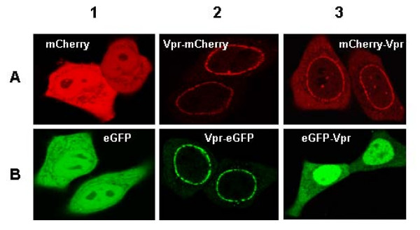
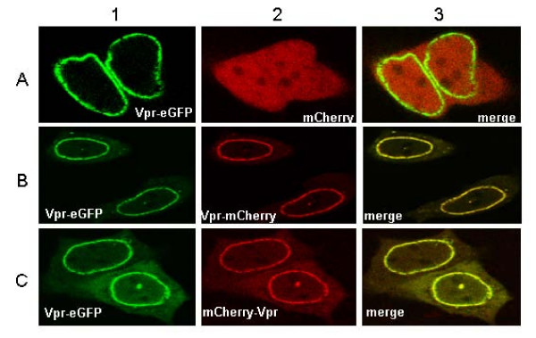
The human immunodeficiency virus type 1 (HIV-1) encodes several regulatory proteins, notably Vpr which influences the survival of the infected cells by causing a G2/M arrest and apoptosis. Such an important role of Vpr in HIV-1 disease progression has fuelled a large number of studies, from its 3D structure to the characterization of specific cellular partners. However, no direct imaging and quantification of Vpr-Vpr interaction in living cells has yet been reported. To address this issue, eGFP- and mCherry proteins were tagged by Vpr, expressed in HeLa cells and their interaction was studied by two photon fluorescence lifetime imaging microscopy and fluorescence correlation spectroscopy.
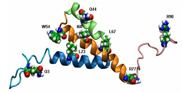
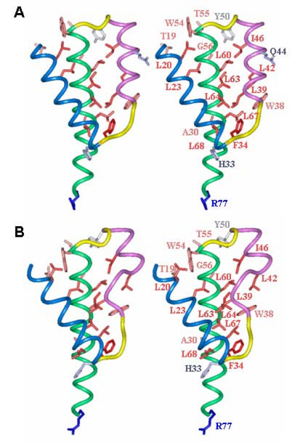
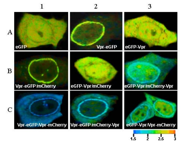
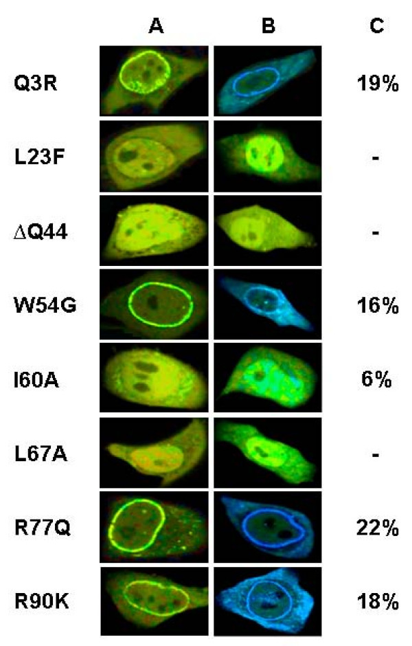
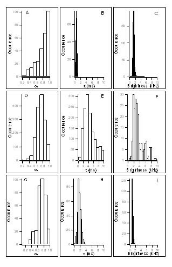
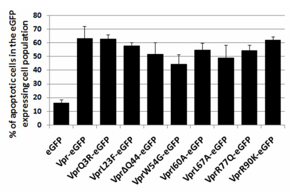
Results show that Vpr forms homo-oligomers at or close to the nuclear envelope. Moreover, Vpr dimers and trimers were found in the cytoplasm and in the nucleus. Point mutations in the three alpha helices of Vpr drastically impaired Vpr oligomerization and localization at the nuclear envelope while point mutations outside the helical regions had no effect. Theoretical structures of Vpr mutants reveal that mutations within the alpha-helices could perturb the leucine zipper like motifs. The DeltaQ44 mutation has the most drastic effect since it likely disrupts the second helix. Finally, all Vpr point mutants caused cell apoptosis suggesting that Vpr-mediated apoptosis functions independently from Vpr oligomerization.
We report that Vpr oligomerization in HeLa cells relies on the hydrophobic core formed by the three alpha helices. This oligomerization is required for Vpr localization at the nuclear envelope but not for Vpr-mediated apoptosis.
人类免疫缺陷病毒1型(HIV-1)编码多种调节蛋白,特别是Vpr,它通过导致G2/M期阻滞和细胞凋亡来影响受感染细胞的存活。Vpr在HIV-1疾病进展中的这一重要作用推动了大量研究,从其三维结构到特定细胞伴侣的表征。然而,尚未有关于活细胞中Vpr-Vpr相互作用的直接成像和定量报道。为了解决这个问题,Vpr标记了eGFP和mCherry蛋白,在HeLa细胞中表达,并通过双光子荧光寿命成像显微镜和荧光相关光谱研究它们的相互作用。
结果表明,Vpr在核膜处或其附近形成同源寡聚体。此外,在细胞质和细胞核中发现了Vpr二聚体和三聚体。Vpr的三个α螺旋中的点突变极大地损害了Vpr的寡聚化和在核膜处的定位,而螺旋区域外的点突变则没有影响。Vpr突变体的理论结构表明,α螺旋内的突变可能会扰乱亮氨酸拉链样基序。DeltaQ44突变的影响最为显著,因为它可能破坏了第二个螺旋。最后,所有Vpr点突变体都导致细胞凋亡,这表明Vpr介导的细胞凋亡功能独立于Vpr寡聚化。
我们报道HeLa细胞中的Vpr寡聚化依赖于由三个α螺旋形成的疏水核心。这种寡聚化是Vpr定位在核膜所必需的,但不是Vpr介导的细胞凋亡所必需的。