• 文献检索
  • 文档翻译
  • 深度研究
  • 学术资讯
  • Suppr Zotero 插件Zotero 插件
  • 邀请有礼
  • 套餐&价格
  • 历史记录
应用&插件
Suppr Zotero 插件Zotero 插件浏览器插件Mac 客户端Windows 客户端微信小程序
定价
高级版会员购买积分包购买API积分包
服务
文献检索文档翻译深度研究API 文档MCP 服务
关于我们
关于 Suppr公司介绍联系我们用户协议隐私条款
关注我们

Suppr 超能文献

核心技术专利:CN118964589B侵权必究
粤ICP备2023148730 号-1Suppr @ 2026

文献检索

告别复杂PubMed语法,用中文像聊天一样搜索,搜遍4000万医学文献。AI智能推荐,让科研检索更轻松。

立即免费搜索

文件翻译

保留排版,准确专业,支持PDF/Word/PPT等文件格式,支持 12+语言互译。

免费翻译文档

深度研究

AI帮你快速写综述,25分钟生成高质量综述,智能提取关键信息,辅助科研写作。

立即免费体验

带蒂子宫平滑肌瘤酷似腹部肿块:一例报告

Pedunculated uterine leiomyoma mimicking abdominal mass: a case report.

作者信息

Spanoudaki Athina, Oikonomou Anastasia, Dimitrova Krasimira, Prassopoulos Panos

机构信息

Department of Radiology, University Hospital of Alexandroupolis, Democritus University of Thrace, Greece.

出版信息

Cases J. 2008 Nov 17;1(1):315. doi: 10.1186/1757-1626-1-315.

DOI:10.1186/1757-1626-1-315
PMID:19014628
原文链接:https://pmc.ncbi.nlm.nih.gov/articles/PMC2588583/
Abstract

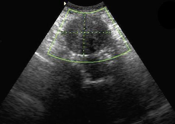
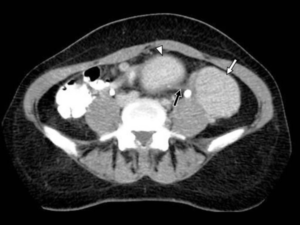
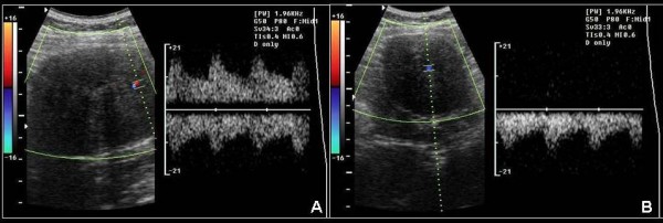
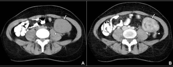
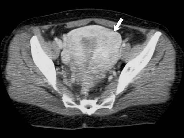
A 48-year-old woman presented with abdominal fullness and a palpable "mass" in the left lower quadrant. Ultrasonography showed a large, rounded, hypoechoic mass. Contrast-enhanced helical CT of the abdomen demonstrated a well-circumscribed, heterogeneously but vividly enhancing mass. The uterus had a leiomyomatous configuration on CT. Uterus and mass revealed the same enhancing pattern. Thin section CT revealed a long, thin stalk connecting the mass with the body of the uterus. Surgical removal of both uterus and the mass confirmed the diagnosis of a pedunculated subserosal leiomyoma originating from a leiomyomatous uterus.

摘要

一名48岁女性因腹部胀满及左下腹可触及“肿块”就诊。超声检查显示一个大的、圆形的低回声肿块。腹部增强螺旋CT显示一个边界清晰、不均匀但强化明显的肿块。CT上子宫呈平滑肌瘤形态。子宫和肿块显示出相同的强化模式。薄层CT显示一条细长的蒂将肿块与子宫体相连。手术切除子宫和肿块证实诊断为起源于平滑肌瘤性子宫的带蒂浆膜下平滑肌瘤。

https://cdn.ncbi.nlm.nih.gov/pmc/blobs/09eb/2588583/e71bd776a2b2/1757-1626-1-315-5.jpg
https://cdn.ncbi.nlm.nih.gov/pmc/blobs/09eb/2588583/112174ab3d13/1757-1626-1-315-1.jpg
https://cdn.ncbi.nlm.nih.gov/pmc/blobs/09eb/2588583/ae094aabf620/1757-1626-1-315-2.jpg
https://cdn.ncbi.nlm.nih.gov/pmc/blobs/09eb/2588583/04936e855f15/1757-1626-1-315-3.jpg
https://cdn.ncbi.nlm.nih.gov/pmc/blobs/09eb/2588583/0c0854b15887/1757-1626-1-315-4.jpg
https://cdn.ncbi.nlm.nih.gov/pmc/blobs/09eb/2588583/e71bd776a2b2/1757-1626-1-315-5.jpg

相似文献

1
Pedunculated uterine leiomyoma mimicking abdominal mass: a case report.带蒂子宫平滑肌瘤酷似腹部肿块:一例报告
Cases J. 2008 Nov 17;1(1):315. doi: 10.1186/1757-1626-1-315.
2
Large subserous uterine leiomyoma presenting as intraabdominal tumor: A case report.表现为腹腔内肿瘤的巨大浆膜下子宫平滑肌瘤:病例报告
Rare Tumors. 2024 Sep 12;16:20363613241285089. doi: 10.1177/20363613241285089. eCollection 2024.
3
Emergency Surgical Treatment of a Large Pedunculated Subserosal Uterine Leiomyoma With Torsion: A Rare Cause of Acute Abdomen.带蒂浆膜下子宫平滑肌瘤扭转的急诊手术治疗:急性腹痛的罕见原因
Cureus. 2024 Jan 11;16(1):e52136. doi: 10.7759/cureus.52136. eCollection 2024 Jan.
4
A subserosal, pedunculated, multilocular uterine leiomyoma with ovarian tumor-like morphology and histological architecture of adenomatoid tumors: a case report and review of the literature.一例具有腺瘤样瘤的卵巢肿瘤样形态和组织学结构的浆膜下带蒂多房性子宫平滑肌瘤:病例报告及文献复习
J Med Case Rep. 2016 Dec 20;10(1):352. doi: 10.1186/s13256-016-1167-1.
5
An unusual case of pedunculated subserosal leiomyosarcoma of the uterus mimicking ovarian carcinoma.一例罕见的带蒂子宫浆膜下平滑肌肉瘤,酷似卵巢癌。
J Ovarian Res. 2016 Feb 9;9:2. doi: 10.1186/s13048-016-0212-4.
6
Outcomes after uterine artery embolization for pedunculated subserosal leiomyomas.有蒂浆膜下平滑肌瘤子宫动脉栓塞术后的结局
J Vasc Interv Radiol. 2008 May;19(5):657-61. doi: 10.1016/j.jvir.2007.11.022. Epub 2008 Mar 17.
7
A large pedunculated leiomyoma with two-sided cystic degenerations mimicking a bilateral ovarian malignancy: a case report.一例带蒂大平滑肌瘤伴双侧囊性变酷似双侧卵巢恶性肿瘤的病例报告
Eur J Gynaecol Oncol. 2014;35(2):192-4.
8
A case of cystic leiomyoma mimicking an ovarian malignancy.一例酷似卵巢恶性肿瘤的囊性平滑肌瘤病例。
Ann Acad Med Singap. 2004 May;33(3):371-4.
9
Torsion of a Pedunculated Subserosal Leiomyoma: A Rare Cause of Acute Abdominal Pain.带蒂浆膜下平滑肌瘤扭转:急性腹痛的罕见原因。
Cureus. 2023 Nov 6;15(11):e48414. doi: 10.7759/cureus.48414. eCollection 2023 Nov.
10
Uterine artery embolization for pedunculated subserosal fibroids.子宫动脉栓塞术治疗有蒂浆膜下子宫肌瘤
AJR Am J Roentgenol. 2005 Feb;184(2):399-402. doi: 10.2214/ajr.184.2.01840399.

引用本文的文献

1
Large subserous uterine leiomyoma presenting as intraabdominal tumor: A case report.表现为腹腔内肿瘤的巨大浆膜下子宫平滑肌瘤:病例报告
Rare Tumors. 2024 Sep 12;16:20363613241285089. doi: 10.1177/20363613241285089. eCollection 2024.
2
The Differential Diagnosis of Abdominal Mass: A Case of Uterine Leiomyoma.腹部肿块的鉴别诊断:一例子宫平滑肌瘤病例
Cureus. 2024 Jul 22;16(7):e65126. doi: 10.7759/cureus.65126. eCollection 2024 Jul.
3
Massive uterine fibroid: a diagnostic dilemma: a case report and review of the literature.巨大子宫肌瘤:诊断难题——病例报告及文献复习。

本文引用的文献

1
Uterine smooth-muscle tumors with unusual growth patterns: imaging with pathologic correlation.具有异常生长模式的子宫平滑肌瘤:影像学与病理对照
AJR Am J Roentgenol. 2007 Jan;188(1):246-55. doi: 10.2214/AJR.05.1070.
2
Answer to case of the month #103. Large subserosal uterine leiomyoma with cystic degeneration presenting as an abdominal mass.第103期月度病例解答。巨大浆膜下子宫平滑肌瘤伴囊性变,表现为腹部肿块。
Can Assoc Radiol J. 2005 Oct;56(4):245-7.
3
Diffusely enlarged uterus: evaluation with MR imaging.
J Med Case Rep. 2021 Jul 13;15(1):344. doi: 10.1186/s13256-021-02959-3.
4
Dynamics of urinary oxidative stress biomarkers: 8-hydroxy-2'-deoxyguanosine and 8-isoprostane in uterine leiomyomas.子宫肌瘤中尿氧化应激生物标志物的动态变化:8-羟基-2'-脱氧鸟苷和8-异前列腺素
J Midlife Health. 2016 Jan-Mar;7(1):8-14. doi: 10.4103/0976-7800.179173.
Radiographics. 2003 Nov-Dec;23(6):1423-39. doi: 10.1148/rg.236035033.
4
Unusual appearances of uterine leiomyomas: MR imaging findings and their histopathologic backgrounds.子宫平滑肌瘤的不寻常表现:磁共振成像结果及其组织病理学背景
Radiographics. 1999 Oct;19 Spec No:S131-45. doi: 10.1148/radiographics.19.suppl_1.g99oc04s131.
5
Uterine leiomyomas: histopathologic features, MR imaging findings, differential diagnosis, and treatment.子宫平滑肌瘤:组织病理学特征、磁共振成像表现、鉴别诊断及治疗
Radiographics. 1999 Sep-Oct;19(5):1179-97. doi: 10.1148/radiographics.19.5.g99se131179.
6
Red degeneration of uterine leiomyoma: MR appearance.子宫平滑肌瘤红色变性:磁共振成像表现
J Comput Assist Tomogr. 1994 Nov-Dec;18(6):925-8. doi: 10.1097/00004728-199411000-00014.
7
Uterine leiomyomas: correlation of MR, histopathologic findings, and symptoms.子宫平滑肌瘤:磁共振成像、组织病理学表现及症状的相关性
Radiology. 1986 Feb;158(2):385-91. doi: 10.1148/radiology.158.2.3753623.
8
CT appearance of uterine leiomyomas.子宫平滑肌瘤的CT表现。
Radiographics. 1990 Nov;10(6):999-1007. doi: 10.1148/radiographics.10.6.2259770.
9
Imaging of uterine leiomyomas.
AJR Am J Roentgenol. 1992 Apr;158(4):799-805. doi: 10.2214/ajr.158.4.1546595.