• 文献检索
  • 文档翻译
  • 深度研究
  • 学术资讯
  • Suppr Zotero 插件Zotero 插件
  • 邀请有礼
  • 套餐&价格
  • 历史记录
应用&插件
Suppr Zotero 插件Zotero 插件浏览器插件Mac 客户端Windows 客户端微信小程序
定价
高级版会员购买积分包购买API积分包
服务
文献检索文档翻译深度研究API 文档MCP 服务
关于我们
关于 Suppr公司介绍联系我们用户协议隐私条款
关注我们

Suppr 超能文献

核心技术专利:CN118964589B侵权必究
粤ICP备2023148730 号-1Suppr @ 2026

文献检索

告别复杂PubMed语法,用中文像聊天一样搜索,搜遍4000万医学文献。AI智能推荐,让科研检索更轻松。

立即免费搜索

文件翻译

保留排版,准确专业,支持PDF/Word/PPT等文件格式,支持 12+语言互译。

免费翻译文档

深度研究

AI帮你快速写综述,25分钟生成高质量综述,智能提取关键信息,辅助科研写作。

立即免费体验

一个尾侧增殖生长中心对正在形成的心管的两极都有贡献。

A caudal proliferating growth center contributes to both poles of the forming heart tube.

作者信息

van den Berg Gert, Abu-Issa Radwan, de Boer Bouke A, Hutson Mary R, de Boer Piet A J, Soufan Alexandre T, Ruijter Jan M, Kirby Margaret L, van den Hoff Maurice J B, Moorman Antoon F M

机构信息

Heart Failure Research Center, Academic Medical Center, Amsterdam, The Netherlands.

出版信息

Circ Res. 2009 Jan 30;104(2):179-88. doi: 10.1161/CIRCRESAHA.108.185843. Epub 2008 Dec 4.

DOI:10.1161/CIRCRESAHA.108.185843
PMID:19059840
原文链接:https://pmc.ncbi.nlm.nih.gov/articles/PMC2683147/
Abstract

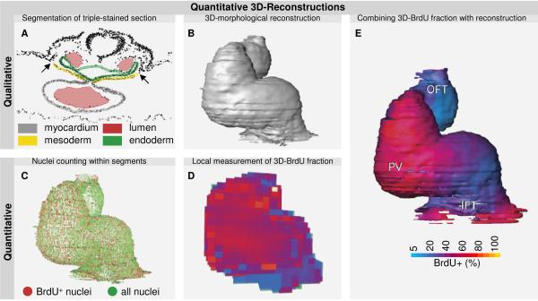
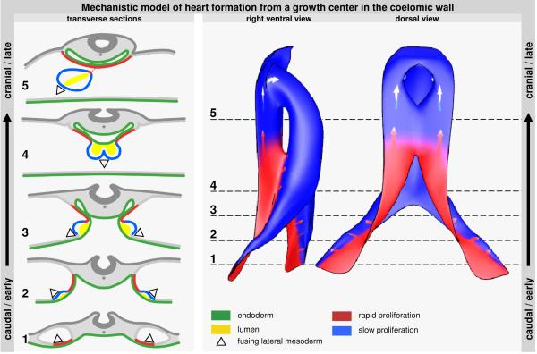
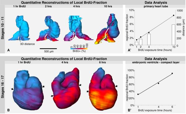
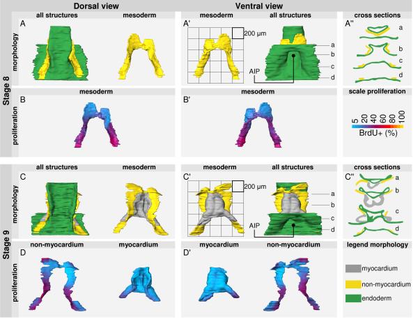
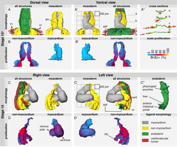
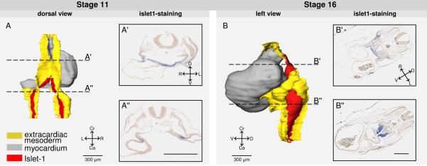
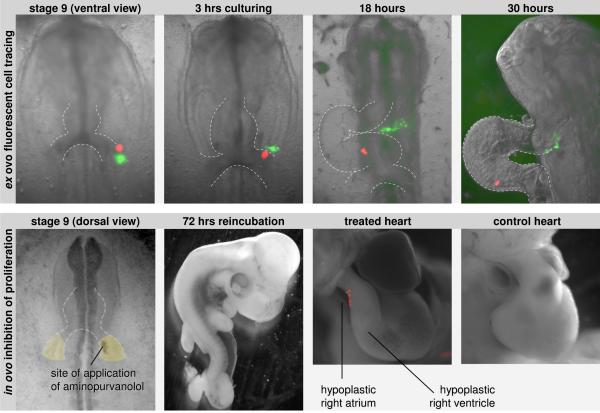
Recent studies have shown that the primary heart tube continues to grow by addition of cells from the coelomic wall. This growth occurs concomitantly with embryonic folding and formation of the coelomic cavity, making early heart formation morphologically complex. A scarcity of data on localized growth parameters further hampers the understanding of cardiac growth. Therefore, we investigated local proliferation during early heart formation. Firstly, we determined the cell cycle length of primary myocardium of the early heart tube to be 5.5 days, showing that this myocardium is nonproliferating and implying that initial heart formation occurs solely by addition of cells. In line with this, we show that the heart tube rapidly lengthens at its inflow by differentiation of recently divided precursor cells. To track the origin of these cells, we made quantitative 3D reconstructions of proliferation in the forming heart tube and the mesoderm of its flanking coelomic walls. These reconstructions show a single, albeit bilateral, center of rapid proliferation in the caudomedial pericardial back wall. This center expresses Islet1. Cell tracing showed that cells from this caudal growth center, besides feeding into the venous pole of the heart, also move cranially via the dorsal pericardial mesoderm and differentiate into myocardium at the arterial pole. Inhibition of caudal proliferation impairs the formation of both the atria and the right ventricle. These data show how a proliferating growth center in the caudal coelomic wall elongates the heart tube at both its venous and arterial pole, providing a morphological mechanism for early heart formation.

摘要

最近的研究表明,原始心管通过添加来自体腔壁的细胞而持续生长。这种生长与胚胎折叠和体腔的形成同时发生,使得早期心脏形成在形态学上变得复杂。关于局部生长参数的数据匮乏进一步阻碍了对心脏生长的理解。因此,我们研究了早期心脏形成过程中的局部增殖情况。首先,我们确定早期心管的原始心肌细胞周期长度为5.5天,这表明该心肌不增殖,意味着最初的心脏形成仅通过细胞添加来实现。与此一致的是,我们发现心管在其流入端通过最近分裂的前体细胞的分化而迅速延长。为了追踪这些细胞的起源,我们对正在形成的心管及其侧翼体腔壁的中胚层中的增殖情况进行了定量三维重建。这些重建显示在尾内侧心包后壁有一个单一的(尽管是双侧的)快速增殖中心。这个中心表达Islet1。细胞追踪显示,来自这个尾侧生长中心的细胞,除了进入心脏的静脉极外,还通过背侧心包中胚层向头侧移动,并在动脉极分化为心肌。抑制尾侧增殖会损害心房和右心室的形成。这些数据表明尾侧体腔壁中的一个增殖生长中心如何在心脏管的静脉极和动脉极使其延长,为早期心脏形成提供了一种形态学机制。

https://cdn.ncbi.nlm.nih.gov/pmc/blobs/a786/2683147/011c8bd59860/nihms-109705-f0007.jpg
https://cdn.ncbi.nlm.nih.gov/pmc/blobs/a786/2683147/5692052982ab/nihms-109705-f0001.jpg
https://cdn.ncbi.nlm.nih.gov/pmc/blobs/a786/2683147/2690f05279ac/nihms-109705-f0002.jpg
https://cdn.ncbi.nlm.nih.gov/pmc/blobs/a786/2683147/d93f2b111518/nihms-109705-f0003.jpg
https://cdn.ncbi.nlm.nih.gov/pmc/blobs/a786/2683147/21f2e93b9d86/nihms-109705-f0004.jpg
https://cdn.ncbi.nlm.nih.gov/pmc/blobs/a786/2683147/60219078dc27/nihms-109705-f0005.jpg
https://cdn.ncbi.nlm.nih.gov/pmc/blobs/a786/2683147/61e1934daf37/nihms-109705-f0006.jpg
https://cdn.ncbi.nlm.nih.gov/pmc/blobs/a786/2683147/011c8bd59860/nihms-109705-f0007.jpg

相似文献

1
A caudal proliferating growth center contributes to both poles of the forming heart tube.一个尾侧增殖生长中心对正在形成的心管的两极都有贡献。
Circ Res. 2009 Jan 30;104(2):179-88. doi: 10.1161/CIRCRESAHA.108.185843. Epub 2008 Dec 4.
2
Conotruncal myocardium arises from a secondary heart field.圆锥干心肌起源于第二心脏场。
Development. 2001 Aug;128(16):3179-88. doi: 10.1242/dev.128.16.3179.
3
Regionalized sequence of myocardial cell growth and proliferation characterizes early chamber formation.心肌细胞生长和增殖的区域化序列是早期心室形成的特征。
Circ Res. 2006 Sep 1;99(5):545-52. doi: 10.1161/01.RES.0000239407.45137.97. Epub 2006 Aug 3.
4
Epicardium and myocardium separate from a common precursor pool by crosstalk between bone morphogenetic protein- and fibroblast growth factor-signaling pathways.心外膜和心肌通过骨形态发生蛋白信号通路与成纤维细胞生长因子信号通路之间的相互作用,从共同的前体细胞库中分离出来。
Circ Res. 2009 Aug 28;105(5):431-41. doi: 10.1161/CIRCRESAHA.109.203083. Epub 2009 Jul 23.
5
Induction of the avian coelom with associated vitelline blood circulation by Rauber's sickle derived junctional endoblast and its fundamental role in heart formation.劳伯氏镰刀衍生的连接内胚层诱导鸟类体腔及其相关的卵黄血液循环及其在心脏形成中的基本作用。
J Morphol. 2004 Jan;259(1):21-32. doi: 10.1002/jmor.10152.
6
The sinus venosus progenitors separate and diversify from the first and second heart fields early in development.窦房静脉祖细胞在发育早期从第一和第二心区分离并多样化。
Cardiovasc Res. 2010 Jul 1;87(1):92-101. doi: 10.1093/cvr/cvq033. Epub 2010 Jan 28.
7
Growth of the developing mouse heart: an interactive qualitative and quantitative 3D atlas.发育中老鼠心脏的生长:一个交互式的定性和定量 3D 图谱。
Dev Biol. 2012 Aug 15;368(2):203-13. doi: 10.1016/j.ydbio.2012.05.001. Epub 2012 May 14.
8
Nkx2.5-negative myocardium of the posterior heart field and its correlation with podoplanin expression in cells from the developing cardiac pacemaking and conduction system.后心区的Nkx2.5阴性心肌及其与发育中心脏起搏和传导系统细胞中血小板内皮细胞黏附分子表达的相关性。
Anat Rec (Hoboken). 2007 Jan;290(1):115-22. doi: 10.1002/ar.20406.
9
The outflow tract of the heart is recruited from a novel heart-forming field.心脏的流出道源自一个新的心脏形成区域。
Dev Biol. 2001 Oct 1;238(1):97-109. doi: 10.1006/dbio.2001.0409.
10
BMP and FGF regulate the differentiation of multipotential pericardial mesoderm into the myocardial or epicardial lineage.骨形态发生蛋白(BMP)和成纤维细胞生长因子(FGF)调节多能心包中胚层向心肌或心外膜谱系的分化。
Dev Biol. 2006 Jul 15;295(2):507-22. doi: 10.1016/j.ydbio.2006.03.033. Epub 2006 Apr 3.

引用本文的文献

1
Role of Maternal Obesity in Offspring Cardiovascular Development and Congenital Heart Defects.母体肥胖在子代心血管发育和先天性心脏病中的作用。
J Am Heart Assoc. 2025 May 6;14(9):e039684. doi: 10.1161/JAHA.124.039684. Epub 2025 May 2.
2
The molecular mechanisms of cardiac development and related diseases.心脏发育及相关疾病的分子机制。
Signal Transduct Target Ther. 2024 Dec 23;9(1):368. doi: 10.1038/s41392-024-02069-8.
3
Mesodermal fibronectin controls cell shape, polarity, and mechanotransduction in the second heart field during cardiac outflow tract development.

本文引用的文献

1
A series of normal stages in the development of the chick embryo.鸡胚胎发育的一系列正常阶段。
J Morphol. 1951 Jan;88(1):49-92.
2
Patterning of the heart field in the chick.鸡胚心脏区域的模式形成
Dev Biol. 2008 Jul 15;319(2):223-33. doi: 10.1016/j.ydbio.2008.04.014. Epub 2008 Apr 23.
3
Islet1 cardiovascular progenitors: a single source for heart lineages?胰岛1心血管祖细胞:心脏谱系的单一来源?
中胚层纤连蛋白在心脏流出道发育过程中控制第二心脏场中的细胞形状、极性和机械转导。
Dev Cell. 2025 Jan 6;60(1):62-84.e7. doi: 10.1016/j.devcel.2024.09.017. Epub 2024 Oct 15.
4
miR-1 as a Key Epigenetic Regulator in Early Differentiation of Cardiac Sinoatrial Region.miR-1 作为心脏窦房结早期分化的关键表观遗传调控因子。
Int J Mol Sci. 2024 Jun 15;25(12):6608. doi: 10.3390/ijms25126608.
5
Human Cardiac Development.人类心脏发育
Adv Exp Med Biol. 2024;1441:3-55. doi: 10.1007/978-3-031-44087-8_1.
6
Deletion of Sarcolemmal Membrane-Associated Protein Isoform 3 (SLMAP3) in Cardiac Progenitors Delays Embryonic Growth of Myocardium without Affecting Hippo Pathway.心肌祖细胞中细胞膜相关蛋白同种型 3(SLMAP3)缺失延缓胚胎心肌生长而不影响 Hippo 通路。
Int J Mol Sci. 2024 Mar 1;25(5):2888. doi: 10.3390/ijms25052888.
7
The Tbx20-TLE interaction is essential for the maintenance of the second heart field.Tbx20 与 TLE 的相互作用对于维持第二心脏场至关重要。
Development. 2023 Nov 1;150(21). doi: 10.1242/dev.201677. Epub 2023 Oct 30.
8
Human Heart Morphogenesis: A New Vision Based on In Vivo Labeling and Cell Tracking.人类心脏形态发生:基于体内标记和细胞追踪的新视角。
Life (Basel). 2023 Jan 6;13(1):165. doi: 10.3390/life13010165.
9
The second heart field: the first 20 years.第二个心脏场:最初的 20 年。
Mamm Genome. 2023 Jun;34(2):216-228. doi: 10.1007/s00335-022-09975-8. Epub 2022 Dec 22.
10
The negative regulation of gene expression by microRNAs as key driver of inducers and repressors of cardiomyocyte differentiation.微小 RNA 对基因表达的负调控作为心肌细胞分化诱导物和抑制剂的关键驱动因素。
Clin Sci (Lond). 2022 Aug 31;136(16):1179-1203. doi: 10.1042/CS20220391.
Development. 2008 Jan;135(2):193-205. doi: 10.1242/dev.001883.
4
The amphibian second heart field: Xenopus islet-1 is required for cardiovascular development.两栖动物的第二心脏区域:非洲爪蟾的胰岛-1对心血管发育至关重要。
Dev Biol. 2007 Nov 15;311(2):297-310. doi: 10.1016/j.ydbio.2007.08.004. Epub 2007 Aug 10.
5
The heart-forming fields: one or multiple?心脏形成区域:是一个还是多个?
Philos Trans R Soc Lond B Biol Sci. 2007 Aug 29;362(1484):1257-65. doi: 10.1098/rstb.2007.2113.
6
Three-dimensional measurement and visualization of morphogenesis applied to cardiac embryology.应用于心脏胚胎学的形态发生的三维测量与可视化
J Microsc. 2007 Mar;225(Pt 3):269-74. doi: 10.1111/j.1365-2818.2007.01742.x.
7
An Nkx2-5/Bmp2/Smad1 negative feedback loop controls heart progenitor specification and proliferation.一个Nkx2-5/Bmp2/Smad1负反馈回路控制心脏祖细胞的特化和增殖。
Cell. 2007 Mar 9;128(5):947-59. doi: 10.1016/j.cell.2007.01.042.
8
Morphological and molecular left-right asymmetries in the development of the proepicardium: a comparative analysis on mouse and chick embryos.心外膜原基发育过程中的形态学和分子左右不对称性:小鼠和鸡胚胎的比较分析
Dev Dyn. 2007 Mar;236(3):684-95. doi: 10.1002/dvdy.21065.
9
Regionalized sequence of myocardial cell growth and proliferation characterizes early chamber formation.心肌细胞生长和增殖的区域化序列是早期心室形成的特征。
Circ Res. 2006 Sep 1;99(5):545-52. doi: 10.1161/01.RES.0000239407.45137.97. Epub 2006 Aug 3.
10
Atrial and ventricular myosin heavy-chain expression in the developing chicken heart: strengths and limitations of non-radioactive in situ hybridization.发育中的鸡心脏中房性和室性肌球蛋白重链的表达:非放射性原位杂交的优势与局限
J Histochem Cytochem. 2006 Jun;54(6):649-64. doi: 10.1369/jhc.5A6846.2006. Epub 2006 Feb 6.