Stone Brian S, Zhang Jiangyang, Mack Devin W, Mori Susumu, Martin Lee J, Northington Frances J
Department of Pediatrics, Eudowood Neonatal Pulmonary Division, Neonatal Research Laboratory, Johns Hopkins University School of Medicine, Baltimore, MD 21287, USA.
Ann Neurol. 2008 Nov;64(5):535-46. doi: 10.1002/ana.21517.
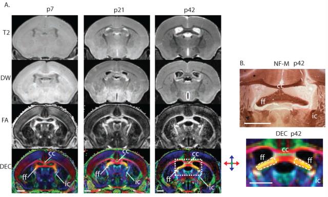
Clinical magnetic resonance studies show delayed and ongoing neurodegeneration after neonatal hypoxia-ischemia (HI), but the mechanisms and timing of this neurodegeneration remain unclear. We used ex vivo diffusion tensor imaging (DTI) and brain neuropathology to determine whether selective injury to white matter tracts occurs after neonatal HI in mice resulting in neural system-associated attrition in remote regions and at delayed times.
The Rice-Vannucci model (unilateral carotid ligation + 45 minutes of hypoxia FiO(2) = 0.08) was used to cause brain injury in postnatal day 7 (p7) C57BL6 mice, and ex vivo DTI and correlative neuropathology were performed at p8, p11, p15, p21, p28, and p42.
DTI provides excellent contrast visualization of unmyelinated white matter in the immature mouse brain. Severe ipsilateral injury to the hippocampus is seen with both histopathology and diffusion-weighted magnetic resonance imaging 24 hours after injury. Injury to axons is evident 24 hours after HI in the hippocampal alveus. By p11 and continuing until p28, the ipsilateral fimbria fornix degenerates. At p15, there is injury and loss of axons entering the ipsilateral septal nucleus followed by ipsilateral septal atrophy. Volume loss in the hippocampus is rapid and severe, but is subacute and significantly slower in the ipsilateral septum. Neonatal HI also interrupts the normal developmental increase in fractional anisotropy in the ipsilateral fimbria but not in the contralateral fimbria from p8 to p42.
In neonatal brain, there is progressive systems-preferential injury after HI. DTI allows unparalleled visualization of this neural network-associated attrition so that it can be followed longitudinally in developing brain.
临床磁共振研究显示新生儿缺氧缺血(HI)后存在延迟性且持续的神经退行性变,但其机制和时间尚不明确。我们运用离体扩散张量成像(DTI)和脑神经病理学方法,来确定新生小鼠HI后白质束是否发生选择性损伤,从而导致远隔区域在延迟时间出现与神经系统相关的萎缩。
采用赖斯-万努奇模型(单侧颈动脉结扎+45分钟低氧,FiO₂ = 0.08)对出生后第7天(p7)的C57BL6小鼠造成脑损伤,并在p8、p11、p15、p21、p28和p42进行离体DTI及相关神经病理学检查。
DTI能出色地显示未成熟小鼠脑内无髓鞘白质的对比影像。损伤后24小时,组织病理学和扩散加权磁共振成像均显示海马同侧有严重损伤。海马齿状回在HI后24小时轴突损伤明显。到p11时,同侧穹窿伞开始退变并持续至p28。在p15时,进入同侧隔核的轴突出现损伤和丢失,随后同侧隔核萎缩。海马体积迅速且严重减小,但同侧隔核的体积减小是亚急性的且明显较慢。从p8到p42,新生HI还会中断同侧穹窿伞中各向异性分数的正常发育性增加,但对侧穹窿伞不受影响。
在新生脑内,HI后存在渐进性的系统优先性损伤。DTI能无与伦比地显示这种与神经网络相关的萎缩,从而可在发育中的脑内进行纵向追踪观察。