Colomiere M, Ward A C, Riley C, Trenerry M K, Cameron-Smith D, Findlay J, Ackland L, Ahmed N
Women's Cancer Research Centre, Royal Women's Hospital, Melbourne, Australia.
Br J Cancer. 2009 Jan 13;100(1):134-44. doi: 10.1038/sj.bjc.6604794. Epub 2008 Dec 16.
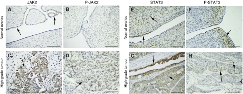
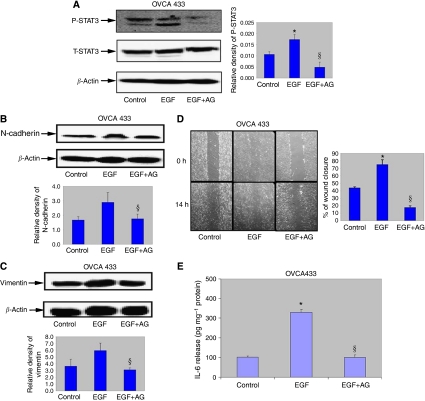
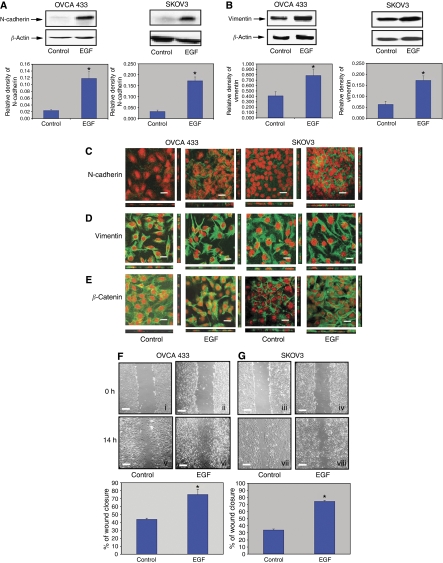
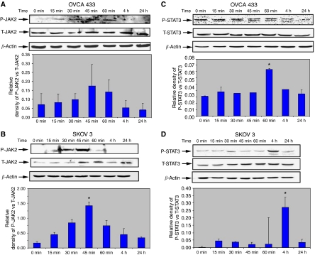
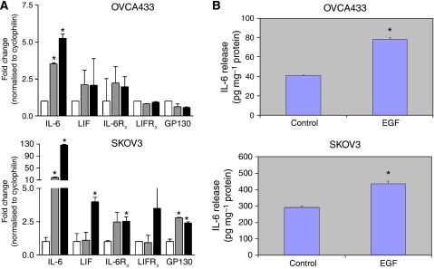
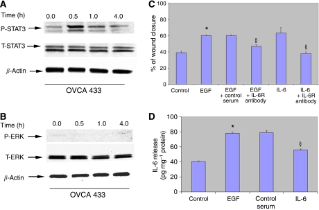
Epidermal growth factor receptor (EGFR) is overexpressed in ovarian carcinomas, with direct or indirect activation of EGFR able to trigger tumour growth. We demonstrate significant activation of both signal transducer and activator of transcription (STAT)3 and its upstream activator Janus kinase (JAK)2, in high-grade ovarian carcinomas compared with normal ovaries and benign tumours. The association between STAT3 activation and migratory phenotype of ovarian cancer cells was investigated by EGF-induced epithelial-mesenchymal transition (EMT) in OVCA 433 and SKOV3 ovarian cancer cell lines. Ligand activation of EGFR induced a fibroblast-like morphology and migratory phenotype, consistent with the upregulation of mesenchyme-associated N-cadherin, vimentin and nuclear translocation of beta-catenin. This occurred concomitantly with activation of the downstream JAK2/STAT3 pathway. Both cell lines expressed interleukin-6 receptor (IL-6R), and treatment with EGF within 1 h resulted in a several-fold enhancement of mRNA expression of IL-6. Consistent with that, EGF treatment of both OVCA 433 and SKOV3 cell lines resulted in enhanced IL-6 production in the serum-free medium. Exogenous addition of IL-6 to OVCA 433 cells stimulated STAT3 activation and enhanced migration. Blocking antibodies against IL-6R inhibited IL-6 production and EGF- and IL-6-induced migration. Specific inhibition of STAT3 activation by JAK2-specific inhibitor AG490 blocked STAT3 phosphorylation, cell motility, induction of N-cadherin and vimentin expression and IL6 production. These data suggest that the activated status of STAT3 in high-grade ovarian carcinomas may occur directly through activation of EGFR or IL-6R or indirectly through induction of IL-6R signalling. Such activation of STAT3 suggests a rationale for a combination of anti-STAT3 and EGFR/IL-6R therapy to suppress the peritoneal spread of ovarian cancer.
表皮生长因子受体(EGFR)在卵巢癌中过度表达,EGFR的直接或间接激活能够触发肿瘤生长。我们证明,与正常卵巢和良性肿瘤相比,高级别卵巢癌中信号转导和转录激活因子(STAT)3及其上游激活因子Janus激酶(JAK)2均有显著激活。通过在OVCA 433和SKOV3卵巢癌细胞系中由表皮生长因子(EGF)诱导的上皮-间质转化(EMT),研究了STAT3激活与卵巢癌细胞迁移表型之间的关联。EGFR的配体激活诱导了成纤维细胞样形态和迁移表型,这与间充质相关的N-钙黏蛋白、波形蛋白的上调以及β-连环蛋白的核转位一致。这一过程与下游JAK2/STAT3通路的激活同时发生。两种细胞系均表达白细胞介素-6受体(IL-6R),在1小时内用EGF处理导致IL-6的mRNA表达增强数倍。与此一致,用EGF处理OVCA 433和SKOV3细胞系均导致无血清培养基中IL-6产生增加。向OVCA 433细胞中额外添加外源性IL-6刺激了STAT3激活并增强了迁移。针对IL-6R的阻断抗体抑制了IL-6产生以及EGF和IL-6诱导的迁移。JAK2特异性抑制剂AG490对STAT3激活的特异性抑制阻断了STAT3磷酸化、细胞运动性、N-钙黏蛋白和波形蛋白表达的诱导以及IL-6产生。这些数据表明,高级别卵巢癌中STAT3的激活状态可能直接通过EGFR或IL-6R的激活发生,或间接通过诱导IL-6R信号传导发生。STAT3的这种激活为抗STAT3与EGFR/IL-6R联合治疗以抑制卵巢癌腹膜播散提供了理论依据。