Lazic Stanley E
Department of Applied Mathematics and Theoretical Physics, Cambridge Computational Biology Institute, University of Cambridge, Cambridge, UK.
BMC Neurosci. 2009 Jan 15;10:5. doi: 10.1186/1471-2202-10-5.
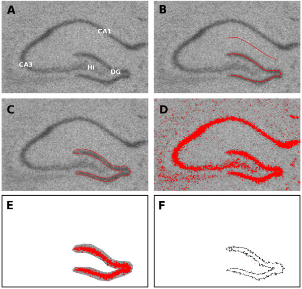
In situ hybridisation (ISH) combined with autoradiography is a standard method of measuring the amount of gene expression in histological sections, but the methods used to quantify gene expression in the resulting digital images vary greatly between studies and can potentially give conflicting results.
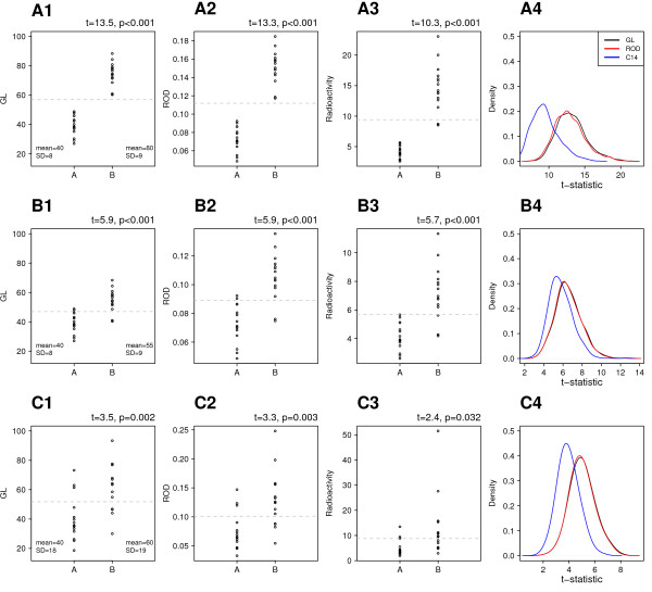
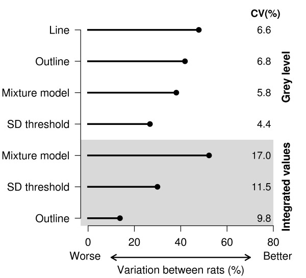
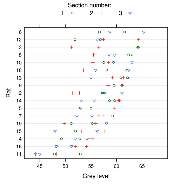
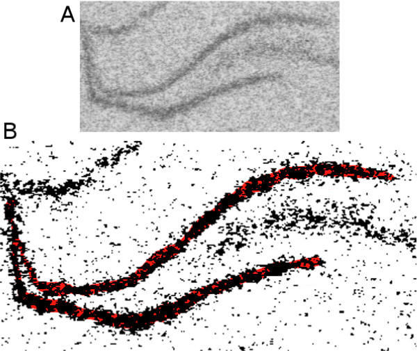
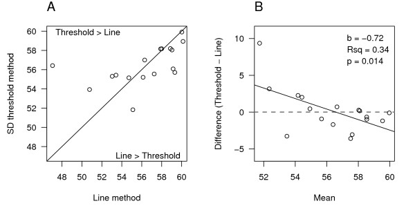
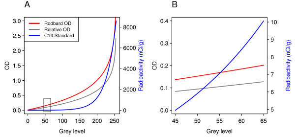
The present study examines commonly used methods for analysing ISH images and demonstrates that these methods are not optimal. Image segmentation based on thresholding can be subject to floor-effects and lead to biased results. In addition, including the area of the structure or region of interest in the calculation of gene expression can lead to a large loss of precision and can also introduce bias. Finally, converting grey level pixel intensities to optical densities or units of radioactivity is unnecessary for most applications and can lead to data with poor statistical properties. A modification of an existing method for selecting the structure or region of interest is introduced which performs better than alternative methods in terms of bias and precision.
Based on these results, suggestions are made to reduce bias, increase precision, and ultimately provide more meaningful results of gene expression data.
原位杂交(ISH)结合放射自显影术是测量组织学切片中基因表达量的标准方法,但在不同研究中,用于量化所得数字图像中基因表达的方法差异很大,且可能得出相互矛盾的结果。
本研究考察了分析ISH图像的常用方法,并证明这些方法并非最优。基于阈值的图像分割可能会受到下限效应的影响,并导致结果有偏差。此外,在基因表达计算中纳入结构或感兴趣区域的面积会导致精度大幅损失,还可能引入偏差。最后,对于大多数应用而言,将灰度像素强度转换为光密度或放射性单位并无必要,且可能导致数据的统计特性不佳。本文介绍了一种对现有选择结构或感兴趣区域方法的改进,该方法在偏差和精度方面比其他方法表现更好。
基于这些结果,提出了减少偏差、提高精度并最终提供更有意义的基因表达数据结果的建议。