Imbalzano Karen M, Tatarkova Iva, Imbalzano Anthony N, Nickerson Jeffrey A
Department of Cell Biology, University of Massachusetts Medical School, Worcester, MA 01655, USA.
Cancer Cell Int. 2009 Mar 16;9:7. doi: 10.1186/1475-2867-9-7.
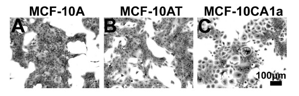
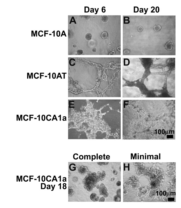
MCF-10A cells are near diploid and normal human mammary epithelial cells. In three-dimensional reconstituted basement membrane culture, they undergo a well-defined program of proliferation, differentiation, and growth arrest, forming acinar structures that recapitulate many aspects of mammary architecture in vivo. The pre-malignant MCF-10AT cells and malignant MCF-10CA1a lines were sequentially derived from the MCF-10A parental cell line first by expression of a constitutively active T24 H-Ras generating the MCF-10AT cell line. This was followed by repeated selection for increasingly aggressive tumor formation from cells recovered from xenograft tumors in immuno-compromised mice, generating the MCF-10CA1a cell line. When inoculated subcutaneously into the flanks of immuno-compromised mice, MCF-10AT cells occasionally form tumors, whereas MCF-10CA1a cells invariably form tumors with a shorter latency than MCF-10AT derived tumors.
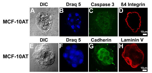
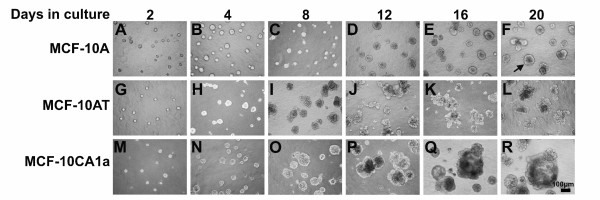
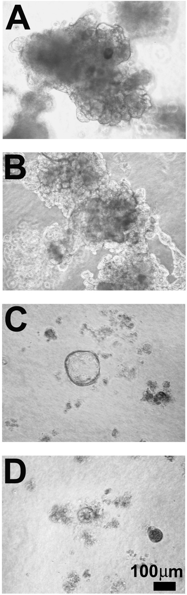
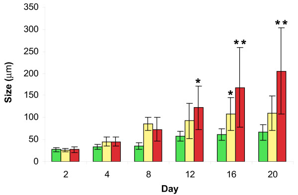
MCF-10AT cells grown in three-dimensional basement membrane culture form complex multi-acinar structures that produce a basement membrane but undergo delayed cell cycle arrest and have incomplete luminal development. MCF-10CA1a cells grown in three-dimensional basement membrane culture form large, hyper-proliferative masses, that retain few characteristics of MCF10A acini and more closely resemble tumors.
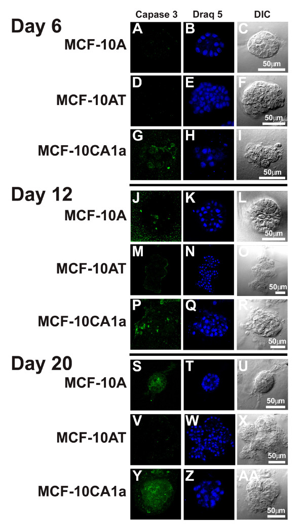
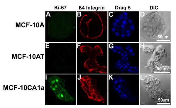
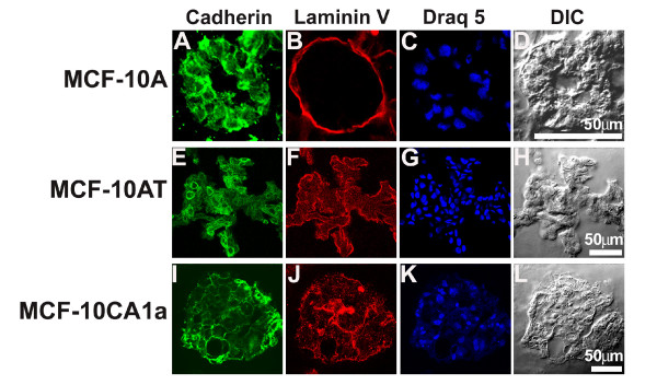
Here we report on the growth and differentiation properties of these three matched cell lines in three-dimensional basement membrane culture. Features of tissue morphogenesis were assessed, including proliferation, basement membrane formation, polarization of alpha-6 beta-4 integrin to the basement membrane, formation of cell:cell junctions, and apoptosis for luminal clearance. The matched series of normal MCF-10A, pre-malignant MCF-10AT, and malignant MCF-10CA1a cells offers a unique opportunity to study the mechanisms of malignant progression both in a three-dimensional microenvironment and in the same cell background.
MCF - 10A细胞是近二倍体的正常人乳腺上皮细胞。在三维重组基底膜培养中,它们经历明确的增殖、分化和生长停滞程序,形成腺泡结构,该结构在体内概括了乳腺结构的许多方面。癌前MCF - 10AT细胞系和恶性MCF - 10CA1a细胞系是首先通过表达组成型活性T24 H - Ras从MCF - 10A亲本细胞系依次衍生而来,从而产生MCF - 10AT细胞系。随后,从免疫缺陷小鼠的异种移植肿瘤中回收的细胞中反复选择形成侵袭性越来越强的肿瘤,从而产生MCF - 10CA1a细胞系。当皮下接种到免疫缺陷小鼠的侧腹时,MCF - 10AT细胞偶尔会形成肿瘤,而MCF - 10CA1a细胞总是会形成肿瘤,且潜伏期比MCF - 10AT衍生的肿瘤短。
在三维基底膜培养中生长的MCF - 10AT细胞形成复杂的多腺泡结构,该结构产生基底膜,但细胞周期停滞延迟且管腔发育不完全。在三维基底膜培养中生长的MCF - 10CA1a细胞形成大的、过度增殖的团块,几乎不保留MCF10A腺泡的特征,更类似于肿瘤。
在此我们报告这三种匹配细胞系在三维基底膜培养中的生长和分化特性。评估了组织形态发生的特征,包括增殖、基底膜形成、α - 6β - 4整合素向基底膜的极化、细胞间连接的形成以及管腔清除的凋亡。正常MCF - 10A、癌前MCF - 10AT和恶性MCF - 10CA1a细胞的匹配系列为在三维微环境和相同细胞背景下研究恶性进展机制提供了独特的机会。