Centelles Miguel N, Qian Cheng, Campanero Miguel A, Irache Juan M
Centro Galénico, Departamento Farmacia y Tecnología Farmacéutica, University of Navarra, Pamplona, Spain.
Int J Nanomedicine. 2008;3(4):451-60. doi: 10.2147/ijn.s3445.
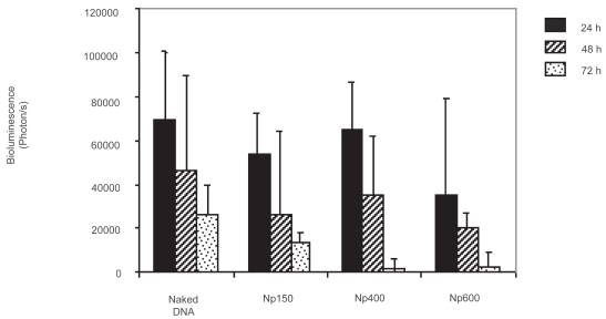
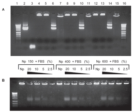
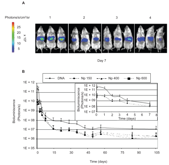
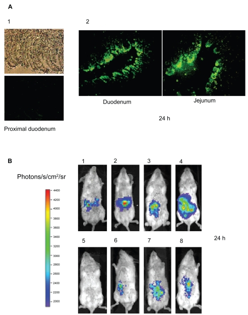
In this work three DNA-chitosan nanoparticle formulations (Np), differing in the molecular weight (MW; 150 kDa, 400 kDa, and 600 kDa) of the polysaccharide, were prepared and administered by two different administration routes: the hydrodynamics-based procedure and the intraduodenal injection. After the hydrodynamic injection, DNA-chitosan nanoparticles were predominantly accumulated in the liver, where the transgene was expressed during at least 105 days. No significant influence of MW was observed on the levels of luciferase expression. The curves of bioluminescence versus time obtained using the charge-coupled device (CCD) camera were described and divided in three phases: (i) the initial phase, (ii) the sustained release step and (iii) the decline phase (promotor inactivation, immunological and physiological processes). From these curves, which describe the transgene expression profile, the behavior of the different formulations as gene delivery systems was characterized. Therefore, the following parameters such as C(max) (maximum level of detected bioluminescence), AUC (area under the bioluminescence-time curve) and MET (mean time of the transgene expression) were calculated. This approach offers the possibility of studying and comparing transgene expression kinetics among a wide variety of gene delivery systems. Finally, the intraduodenal administration of naked DNA permitted the gene transfer in a dose dependent manner quantifiable with the CCD camera within 3 days. Nevertheless, the same administration procedure of the three formulations did not improve the levels of transgene expression obtained with naked DNA. This fact could be explained by the rapid physiological turn-over of enterocytes and by the ability of chitosan nanoparticles to control the DNA release.
在本研究中,制备了三种DNA-壳聚糖纳米颗粒制剂(Np),其多糖的分子量(MW;150 kDa、400 kDa和600 kDa)不同,并通过两种不同的给药途径给药:基于流体动力学的方法和十二指肠内注射。流体动力学注射后,DNA-壳聚糖纳米颗粒主要积聚在肝脏中,转基因在肝脏中至少表达105天。未观察到分子量对荧光素酶表达水平有显著影响。描述了使用电荷耦合器件(CCD)相机获得的生物发光随时间的曲线,并将其分为三个阶段:(i)初始阶段,(ii)持续释放阶段和(iii)下降阶段(启动子失活、免疫和生理过程)。从这些描述转基因表达谱的曲线中,表征了不同制剂作为基因递送系统的行为。因此,计算了以下参数,如C(max)(检测到的生物发光的最大水平)、AUC(生物发光-时间曲线下的面积)和MET(转基因表达的平均时间)。这种方法提供了研究和比较多种基因递送系统中转基因表达动力学的可能性。最后,裸DNA的十二指肠内给药允许以剂量依赖性方式进行基因转移,在3天内可用CCD相机定量。然而,三种制剂的相同给药程序并未提高裸DNA获得的转基因表达水平。这一事实可以通过肠上皮细胞快速的生理更新以及壳聚糖纳米颗粒控制DNA释放的能力来解释。