Bloquel C, Trollet C, Pradines E, Seguin J, Scherman D, Bureau M F
Inserm, U640, Paris, F-75006 France.
BMC Biotechnol. 2006 Mar 8;6:16. doi: 10.1186/1472-6750-6-16.
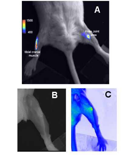
Optical imaging is an attractive non-invasive way to evaluate the expression of a transferred DNA, mainly thanks to its lower cost and ease of realization. In this study optical imaging was evaluated for monitoring and quantification of the mouse knee joint and tibial cranial muscle electrotransfer of a luciferase encoding plasmid. Optical imaging was applied to study the kinetics of luciferase expression in both tissues.
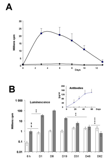
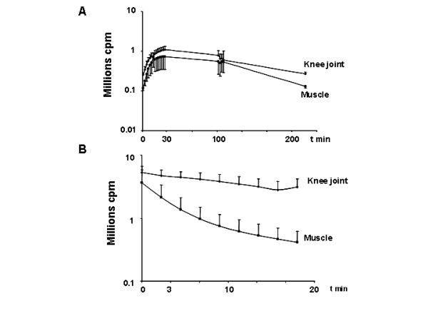
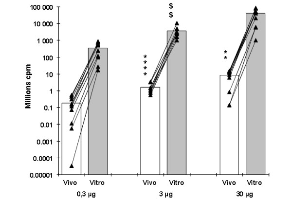
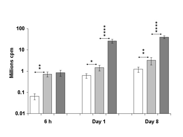
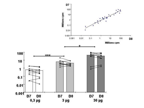
The substrate of luciferase (luciferin) was injected either intraperitonealy (i.p.) or in situ into the muscle or the knee joint. Luminescence resulting from the luciferase-luciferin reaction was measured in vivo with a cooled CCD camera and/or in vitro on tissue lysate. Maximal luminescence of the knee joint and muscle after i.p. (2.5 mg) or local injection of luciferin (50 microg in the knee joint, 100 microg in the muscle) were highly correlated. With the local injection procedure adopted, in vivo and in vitro luminescences measured on the same muscles significantly correlated. Luminescence measurements were reproducible and the signal level was proportional to the amount of plasmid injected. In vivo luciferase activity in the electrotransfered knee joint was detected for two weeks. Intramuscular electrotransfer of 0.3 or 3 microg of plasmid led to stable luciferase expression for 62 days, whereas injecting 30 microg of plasmid resulted in a drop of luminescence three weeks after electrotransfer. These decreases were partially associated with the development of an immune response.
A particular advantage of the i.p. injection of substrate is a widespread distribution at luciferase production sites. We have also highlighted advantages of local injection as a more sensitive detection method with reduced substrate consumption. Besides, this route of injection is relatively free of uncontrolled parameters, such as diffusion to the target organ, crossing of biological barriers and evidencing variations in local enzymatic kinetics, probably related to the reaction medium in the targeted organ. Optical imaging was shown to be a sensitive and relevant technique to quantify variations of luciferase activity in vivo. Further evaluation of the effective amount of luciferase in a given tissue by in vivo optical imaging relies on conditions of the enzymatic reaction and light absorption and presently requires in vitro calibration for each targeted organ.
光学成像作为一种有吸引力的非侵入性方法,主要因其成本较低且易于实现,可用于评估转入DNA的表达情况。在本研究中,对光学成像用于监测和定量小鼠膝关节及胫骨颅侧肌肉中荧光素酶编码质粒的电转染进行了评估。应用光学成像研究了两种组织中荧光素酶表达的动力学。
荧光素酶的底物(荧光素)通过腹腔内注射(i.p.)或原位注射到肌肉或膝关节中。用冷却的电荷耦合器件(CCD)相机在体内和/或在体外对组织裂解物测量荧光素酶 - 荧光素反应产生的发光。腹腔内注射(2.5 mg)或局部注射荧光素(膝关节50 μg,肌肉100 μg)后膝关节和肌肉的最大发光高度相关。采用局部注射方法时,在相同肌肉上测量的体内和体外发光显著相关。发光测量具有可重复性,且信号水平与注射的质粒量成正比。在电转染的膝关节中检测到体内荧光素酶活性持续两周。肌肉内电转染0.3 μg或3 μg质粒导致荧光素酶稳定表达62天,而注射30 μg质粒导致电转染三周后发光下降。这些下降部分与免疫反应的发展有关。
腹腔内注射底物的一个特别优点是在荧光素酶产生部位广泛分布。我们还强调了局部注射作为一种更灵敏的检测方法且底物消耗减少的优点。此外,这种注射途径相对不受诸如向靶器官扩散、生物屏障穿越以及局部酶动力学变化(可能与靶器官中的反应介质有关)等不受控制参数的影响。光学成像被证明是一种灵敏且相关的技术,可用于定量体内荧光素酶活性的变化。通过体内光学成像进一步评估给定组织中荧光素酶的有效量依赖于酶促反应和光吸收的条件,目前每个靶器官都需要进行体外校准。