• 文献检索
  • 文档翻译
  • 深度研究
  • 学术资讯
  • Suppr Zotero 插件Zotero 插件
  • 邀请有礼
  • 套餐&价格
  • 历史记录
应用&插件
Suppr Zotero 插件Zotero 插件浏览器插件Mac 客户端Windows 客户端微信小程序
定价
高级版会员购买积分包购买API积分包
服务
文献检索文档翻译深度研究API 文档MCP 服务
关于我们
关于 Suppr公司介绍联系我们用户协议隐私条款
关注我们

Suppr 超能文献

核心技术专利:CN118964589B侵权必究
粤ICP备2023148730 号-1Suppr @ 2026

文献检索

告别复杂PubMed语法,用中文像聊天一样搜索,搜遍4000万医学文献。AI智能推荐,让科研检索更轻松。

立即免费搜索

文件翻译

保留排版,准确专业,支持PDF/Word/PPT等文件格式,支持 12+语言互译。

免费翻译文档

深度研究

AI帮你快速写综述,25分钟生成高质量综述,智能提取关键信息,辅助科研写作。

立即免费体验

冠状动脉内光学频域成像的三维冠状动脉显微镜检查

Three-dimensional coronary artery microscopy by intracoronary optical frequency domain imaging.

作者信息

Tearney Guillermo J, Waxman Sergio, Shishkov Milen, Vakoc Benjamin J, Suter Melissa J, Freilich Mark I, Desjardins Adrien E, Oh Wang-Yul, Bartlett Lisa A, Rosenberg Mireille, Bouma Brett E

机构信息

Wellman Center for Photomedicine, Massachusetts General Hospital, Department of Pathology, Harvard Medical School, Boston, Massachusetts 02114, USA.

出版信息

JACC Cardiovasc Imaging. 2008 Nov;1(6):752-61. doi: 10.1016/j.jcmg.2008.06.007.

DOI:10.1016/j.jcmg.2008.06.007
PMID:19356512
原文链接:https://pmc.ncbi.nlm.nih.gov/articles/PMC2852244/
Abstract

OBJECTIVES

We present the first clinical experience with intracoronary optical frequency domain imaging (OFDI) in human patients.

BACKGROUND

Intracoronary optical coherence tomography (OCT) is a catheter-based optical imaging modality that is capable of providing microscopic (approximately 7-microm axial resolution, approximately 30-microm transverse resolution), cross-sectional images of the coronary wall. Although the use of OCT has shown substantial promise for imaging coronary microstructure, blood attenuates the OCT signal, necessitating prolonged, proximal occlusion to screen long arterial segments. OFDI is a second-generation form of OCT that is capable of acquiring images at much higher frame rates. The increased speed of OFDI enables rapid, 3-dimensional imaging of long coronary segments after a brief, nonocclusive saline purge.

METHODS

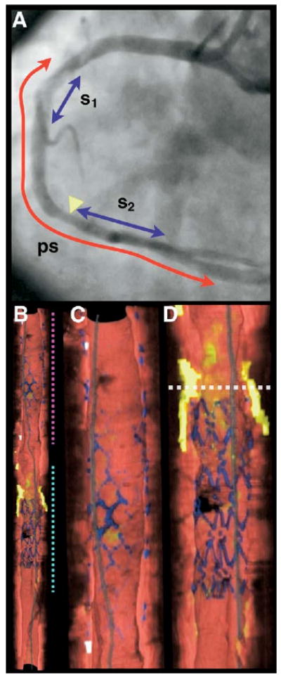
Volumetric OFDI images were obtained in 3 patients after intracoronary stent deployment. Imaging was performed in the left anterior descending and right coronary arteries with the use of a nonocclusive saline purge rates ranging from 3 to 4 ml/s and for purge durations of 3 to 4 s. After imaging, the OFDI datasets were segmented using previously documented criteria and volume rendered.

RESULTS

Good visualization of the artery wall was obtained in all cases, with clear viewing lengths ranging from 3.0 to 7.0 cm at pullback rates ranging from 5 to 20 mm/s. A diverse range of microscopic features were identified in 2 and 3 dimensions, including thin-capped fibroatheromas, calcium, macrophages, cholesterol crystals, bare stent struts, and stents with neointimal hyperplasia. There were no complications of the OFDI procedure.

CONCLUSIONS

Our results demonstrate that OFDI is a viable method for imaging the microstructure of long coronary segments in patients. Given its ability to provide microscopic information in a practical manner, this technology may be useful for studying human coronary pathophysiology in vivo and as a clinical tool for guiding the management of coronary artery disease.

摘要

目的

我们展示了冠状动脉内光学频域成像(OFDI)在人类患者中的首次临床经验。

背景

冠状动脉内光学相干断层扫描(OCT)是一种基于导管的光学成像方式,能够提供冠状动脉壁的微观(轴向分辨率约7微米,横向分辨率约30微米)横断面图像。尽管OCT的应用已显示出在冠状动脉微观结构成像方面的巨大前景,但血液会衰减OCT信号,因此需要长时间的近端闭塞来筛查长动脉段。OFDI是OCT的第二代形式,能够以更高的帧率获取图像。OFDI速度的提高使得在短暂的非闭塞性盐水冲洗后能够对长冠状动脉段进行快速三维成像。

方法

在3例患者冠状动脉内支架置入后获取容积性OFDI图像。使用非闭塞性盐水冲洗,冲洗速度为3至4 ml/s,冲洗持续时间为3至4 s,在左前降支和右冠状动脉进行成像。成像后,根据先前记录的标准对OFDI数据集进行分割并进行容积渲染。

结果

所有病例均获得了动脉壁的良好可视化效果,在5至20 mm/s的回撤速度下,清晰的观察长度为3.0至7.0 cm。在二维和三维中识别出了多种微观特征,包括薄帽纤维粥样斑块、钙、巨噬细胞、胆固醇晶体、裸支架支柱以及伴有内膜增生的支架。OFDI操作无并发症发生。

结论

我们的结果表明,OFDI是一种对患者长冠状动脉段微观结构成像的可行方法。鉴于其能够以实用的方式提供微观信息,该技术可能有助于在体内研究人类冠状动脉病理生理学,并作为指导冠状动脉疾病管理的临床工具。

https://cdn.ncbi.nlm.nih.gov/pmc/blobs/14c5/2852244/c09d177c3d12/nihms185889f7.jpg
https://cdn.ncbi.nlm.nih.gov/pmc/blobs/14c5/2852244/0fbb2b9f0b2f/nihms185889f1.jpg
https://cdn.ncbi.nlm.nih.gov/pmc/blobs/14c5/2852244/1bce9ecab090/nihms185889f2.jpg
https://cdn.ncbi.nlm.nih.gov/pmc/blobs/14c5/2852244/fd056416f82c/nihms185889f3.jpg
https://cdn.ncbi.nlm.nih.gov/pmc/blobs/14c5/2852244/f93593c07cb5/nihms185889f4.jpg
https://cdn.ncbi.nlm.nih.gov/pmc/blobs/14c5/2852244/c656735e5b33/nihms185889f5.jpg
https://cdn.ncbi.nlm.nih.gov/pmc/blobs/14c5/2852244/fe95cb9817c0/nihms185889f6.jpg
https://cdn.ncbi.nlm.nih.gov/pmc/blobs/14c5/2852244/c09d177c3d12/nihms185889f7.jpg

相似文献

1
Three-dimensional coronary artery microscopy by intracoronary optical frequency domain imaging.冠状动脉内光学频域成像的三维冠状动脉显微镜检查
JACC Cardiovasc Imaging. 2008 Nov;1(6):752-61. doi: 10.1016/j.jcmg.2008.06.007.
2
First-in-man evaluation of intravascular optical frequency domain imaging (OFDI) of Terumo: a comparison with intravascular ultrasound and quantitative coronary angiography.首例人体血管内光学频域成像(OFDI)的评估:与血管内超声和定量冠状动脉造影的比较。
EuroIntervention. 2011 Apr;6(9):1037-45. doi: 10.4244/EIJV6I9A182.
3
Coronary optical frequency domain imaging (OFDI) for in vivo evaluation of stent healing: comparison with light and electron microscopy.冠状动脉光频域成像(OFDI)用于评估支架愈合的体内情况:与光镜和电子显微镜的比较。
Eur Heart J. 2010 Jul;31(14):1792-801. doi: 10.1093/eurheartj/ehq168. Epub 2010 Jun 5.
4
Ex vivo assessment of vascular response to coronary stents by optical frequency domain imaging.光学频域成像技术对冠状动脉支架血管反应的离体评估。
JACC Cardiovasc Imaging. 2012 Jan;5(1):71-82. doi: 10.1016/j.jcmg.2011.09.015.
5
Evaluation of the influence of cardiac motion on the accuracy and reproducibility of longitudinal measurements and the corresponding image quality in optical frequency domain imaging: an ex vivo investigation of the optimal pullback speed.评估心脏运动对光学频域成像中纵向测量的准确性和可重复性以及相应图像质量的影响:最佳回撤速度的离体研究。
Int J Cardiovasc Imaging. 2015 Aug;31(6):1115-23. doi: 10.1007/s10554-015-0676-0. Epub 2015 May 14.
6
Recent advances in intracoronary imaging techniques: focus on optical coherence tomography.冠状动脉内成像技术的最新进展:聚焦于光学相干断层扫描
Expert Rev Med Devices. 2008 Nov;5(6):691-7. doi: 10.1586/17434440.5.6.691.
7
Optical coherence tomography in unprotected left main coronary artery stenting.光学相干断层成像术在非保护左主干冠状动脉支架置入术中的应用。
EuroIntervention. 2010 May;6(1):94-9.
8
Intracoronary optical coherence tomography: a comprehensive review clinical and research applications.冠状动脉光学相干断层成像术:全面综述临床和研究应用。
JACC Cardiovasc Interv. 2009 Nov;2(11):1035-46. doi: 10.1016/j.jcin.2009.06.019.
9
Googling the coronary: fiberoptics and a computer provide the answers.谷歌搜索冠状动脉:光纤和计算机给出答案。
JACC Cardiovasc Imaging. 2008 Nov;1(6):762-4. doi: 10.1016/j.jcmg.2008.07.015.
10
Automatic segmentation of in-vivo intra-coronary optical coherence tomography images to assess stent strut apposition and coverage.利用体内冠状动脉光学相干断层扫描图像自动分割评估支架梁贴壁和覆盖率。
Int J Cardiovasc Imaging. 2012 Feb;28(2):229-41. doi: 10.1007/s10554-011-9824-3. Epub 2011 Feb 24.

引用本文的文献

1
Flexible catheter optical coherence tomography of the porcine middle ear via the Eustachian tube using a 3D-printed reflective objective.使用3D打印反射物镜经咽鼓管对猪中耳进行柔性导管光学相干断层扫描。
J Biomed Opt. 2025 Jul;30(7):076002. doi: 10.1117/1.JBO.30.7.076002. Epub 2025 Jul 3.
2
Artificial Intelligence-Based Algorithm for Stent Coverage Assessments.基于人工智能的支架覆盖评估算法
J Pers Med. 2025 Apr 11;15(4):151. doi: 10.3390/jpm15040151.
3
Efficacy and Safety of High-Frequency Optical Coherence Tomography (HF-OCT) for Coronary Imaging: A Multicenter Study.

本文引用的文献

1
Safety and feasibility of a new non-occlusive technique for facilitated intracoronary optical coherence tomography (OCT) acquisition in various clinical and anatomical scenarios.一种新型非闭塞技术用于在各种临床和解剖学场景中辅助进行冠状动脉光学相干断层扫描(OCT)采集的安全性和可行性。
EuroIntervention. 2007 Nov;3(3):365-70. doi: 10.4244/eijv3i3a66.
2
Intravascular optical coherence tomography: optimisation of image acquisition and quantitative assessment of stent strut apposition.血管内光学相干断层扫描:图像采集的优化及支架小梁贴壁的定量评估
EuroIntervention. 2007 May;3(1):128-36.
3
High-speed optical frequency-domain imaging.
高频光学相干断层扫描(HF-OCT)用于冠状动脉成像的有效性和安全性:一项多中心研究。
J Soc Cardiovasc Angiogr Interv. 2025 Mar 18;4(3Part A):102577. doi: 10.1016/j.jscai.2025.102577. eCollection 2025 Mar.
4
Imaging human coronary cholesterol/urate crystals with cross-polarized micro-optical coherence tomography.利用交叉偏振显微光学相干断层扫描对人体冠状动脉胆固醇/尿酸盐结晶进行成像。
Front Cardiovasc Med. 2024 Oct 28;11:1433227. doi: 10.3389/fcvm.2024.1433227. eCollection 2024.
5
Label-free biomedical optical imaging.无标记生物医学光学成像。
Nat Photonics. 2023 Dec;17(12):1031-1041. doi: 10.1038/s41566-023-01299-6. Epub 2023 Nov 16.
6
The Emergence of Nanotechnology in the Prognosis and Treatment of Myocardial Infarctions.纳米技术在心肌梗死预后及治疗中的出现。
Recent Pat Nanotechnol. 2025;19(1):35-55. doi: 10.2174/1872210517666230721123453.
7
Single-input polarization-sensitive optical coherence tomography through a catheter.通过导管的单输入偏振敏感光学相干断层扫描术
Biomed Opt Express. 2023 Aug 10;14(9):4609-4626. doi: 10.1364/BOE.497123. eCollection 2023 Sep 1.
8
The role of invasive and non-invasive imaging technologies and calcium modification therapies in the evaluation and management of coronary artery calcifications.有创和无创成像技术以及钙修饰疗法在冠状动脉钙化评估和管理中的作用。
Front Cardiovasc Med. 2023 Apr 6;10:1133510. doi: 10.3389/fcvm.2023.1133510. eCollection 2023.
9
Automated Coronary Optical Coherence Tomography Feature Extraction with Application to Three-Dimensional Reconstruction.基于冠状动脉光学相干断层成像特征提取的三维重建技术应用。
Tomography. 2022 May 17;8(3):1307-1349. doi: 10.3390/tomography8030108.
10
Optical Coherence Tomography and Coronary Dissection: Precious Tool or Useless Surplus?光学相干断层扫描与冠状动脉夹层:宝贵工具还是无用多余之物?
Front Cardiovasc Med. 2022 Apr 1;9:822998. doi: 10.3389/fcvm.2022.822998. eCollection 2022.
高速光学频域成像。
Opt Express. 2003 Nov 3;11(22):2953-63. doi: 10.1364/oe.11.002953.
4
A pyramid approach to subpixel registration based on intensity.一种基于强度的亚像素配准的金字塔方法。
IEEE Trans Image Process. 1998;7(1):27-41. doi: 10.1109/83.650848.
5
Evaluation of dissection after coronary stent implantation by intravascular optical coherence tomography.血管内光学相干断层扫描评估冠状动脉支架植入术后的夹层情况
Clin Cardiol. 2009 Jul;32(7):E47-8. doi: 10.1002/clc.20173.
6
Relationship between a systemic inflammatory marker, plaque inflammation, and plaque characteristics determined by intravascular optical coherence tomography.一种全身炎症标志物、斑块炎症与通过血管内光学相干断层扫描确定的斑块特征之间的关系。
Arterioscler Thromb Vasc Biol. 2007 Aug;27(8):1820-7. doi: 10.1161/ATVBAHA.107.145987. Epub 2007 May 31.
7
Severe intimal hyperplasia after sirolimus eluting stent deployment: evaluation by optical coherence tomography.西罗莫司洗脱支架植入术后严重内膜增生:光学相干断层扫描评估
Heart. 2007 Jun;93(6):754. doi: 10.1136/hrt.2006.091918.
8
Coronary bioabsorbable magnesium stent: 15-month intravascular ultrasound and optical coherence tomography findings.冠状动脉生物可吸收镁支架:15个月血管内超声和光学相干断层扫描结果。
Eur Heart J. 2007 Oct;28(19):2319. doi: 10.1093/eurheartj/ehm119. Epub 2007 May 7.
9
Pathological correlates of late drug-eluting stent thrombosis: strut coverage as a marker of endothelialization.晚期药物洗脱支架血栓形成的病理相关性:支架小梁覆盖率作为内皮化的标志物
Circulation. 2007 May 8;115(18):2435-41. doi: 10.1161/CIRCULATIONAHA.107.693739. Epub 2007 Apr 16.
10
Evaluation by optical coherence tomography of neointimal coverage of sirolimus-eluting stent three months after implantation.雷帕霉素洗脱支架植入三个月后新生内膜覆盖情况的光学相干断层扫描评估。
Am J Cardiol. 2007 Apr 15;99(8):1033-8. doi: 10.1016/j.amjcard.2006.11.068. Epub 2007 Feb 23.