Reisner Y, Meiry G, Zeevi-Levin N, Barac D Y, Reiter I, Abassi Z, Ziv N, Kostin S, Schaper J, Rosen M R, Binah O
Rappaport Family Institute for Research in the Medical Sciences, Ruth and Bruce Rappaport Faculty of Medicine, Technion-Israel Institute of Technology, Haifa, Israel.
J Cell Mol Med. 2009 Mar;13(3):562-73. doi: 10.1111/j.1582-4934.2008.00361.x.
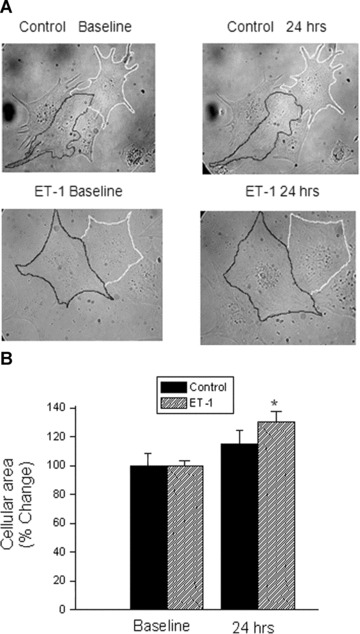
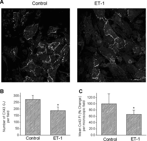
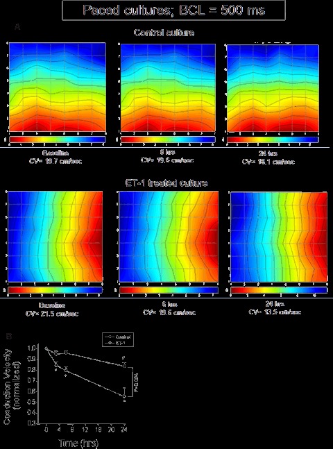
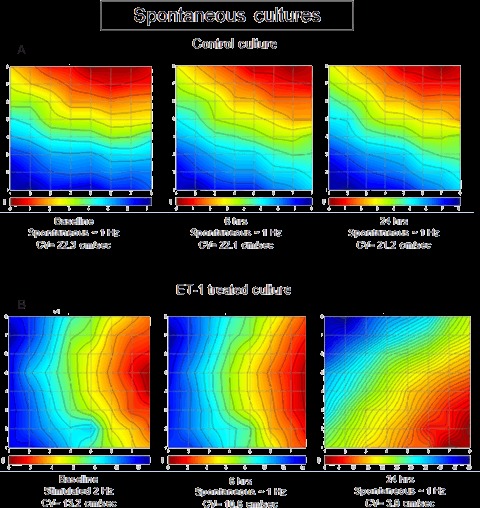
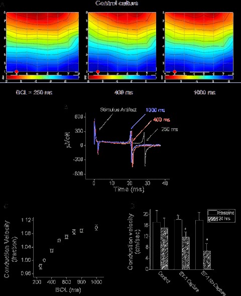
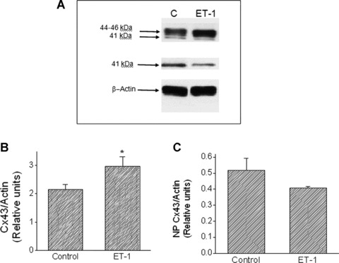
Endothelin-1 (ET-1) is an important contributor to ventricular hypertrophy and failure, which are associated with arrhythmogenesis and sudden death. To elucidate the mechanism(s) underlying the arrhythmogenic effects of ET-1 we tested the hypothesis that long-term (24 hrs) exposure to ET-1 impairs impulse conduction in cultures of neonatal rat ventricular myocytes (NRVM). NRVM were seeded on micro-electrode-arrays (MEAs, Multi Channel Systems, Reutlingen, Germany) and exposed to 50 nM ET-1 for 24 hrs. Hypertrophy was assessed by morphological and molecular methods. Consecutive recordings of paced activation times from the same cultures were conducted at baseline and after 3, 6 and 24 hrs, and activation maps for each time period constructed. Gap junctional Cx43 expression was assessed using Western blot and confocal microscopy of immunofluorescence staining using anti-Cx43 antibodies. ET-1 caused hypertrophy as indicated by a 70% increase in mRNA for atrial natriuretic peptide (P < 0.05), and increased cell areas (P < 0.05) compared to control. ET-1 also caused a time-dependent decrease in conduction velocity that was evident after 3 hrs of exposure to ET-1, and was augmented at 24 hrs, compared to controls (P < 0.01). ET-1 increased total Cx43 protein by approximately 40% (P < 0.05) without affecting non- phosphorylated Cx43 (NP-Cx43) protein expression. Quantitative confocal microscopy showed a approximately 30% decrease in the Cx43 immunofluorescence per field in the ET-1 group (P < 0.05) and a reduced field stain intensity (P < 0.05), compared to controls. ET-1-induced hypertrophy was accompanied by reduction in conduction velocity and gap junctional remodelling. The reduction in conduction velocity may play a role in ET-1 induced susceptibility to arrhythmogenesis.
内皮素-1(ET-1)是导致心室肥厚和衰竭的重要因素,而心室肥厚和衰竭与心律失常及猝死相关。为阐明ET-1致心律失常作用的潜在机制,我们验证了以下假说:长期(24小时)暴露于ET-1会损害新生大鼠心室肌细胞(NRVM)培养物中的冲动传导。将NRVM接种于微电极阵列(MEA,德国罗伊特林根的多通道系统公司)上,并暴露于50 nM的ET-1中24小时。通过形态学和分子方法评估肥厚情况。在基线以及3、6和24小时后,对同一培养物的起搏激活时间进行连续记录,并构建每个时间段的激活图。使用蛋白质免疫印迹法以及使用抗Cx43抗体进行免疫荧光染色的共聚焦显微镜评估缝隙连接蛋白Cx43的表达。与对照组相比,ET-1导致心房利钠肽mRNA增加70%(P<0.05),表明出现肥厚,并且细胞面积增加(P<0.05)。与对照组相比,ET-1还导致传导速度呈时间依赖性下降,在暴露于ET-1 3小时后就很明显,在24小时时进一步增强(P<0.01)。ET-1使总Cx43蛋白增加约40%(P<0.05),但不影响非磷酸化Cx43(NP-Cx43)蛋白表达。定量共聚焦显微镜显示,与对照组相比,ET-1组每视野的Cx43免疫荧光降低约30%(P<0.05),且视野染色强度降低(P<0.05)。ET-1诱导的肥厚伴有传导速度降低和缝隙连接重塑。传导速度降低可能在ET-1诱导的心律失常易感性中起作用。