Saito Anneyuko I, Li Jonathan G, Liu Chihray, Olivier Kenneth R, Dempsey James F
Department of Radiation Oncology, University of Florida, Gainesville, FL, U.S.A.
ViewRay Incorporated, Oakwood Village, OH, U.S.A.
J Appl Clin Med Phys. 2009 Apr 30;10(2):92-103. doi: 10.1120/jacmp.v10i2.2847.
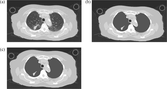
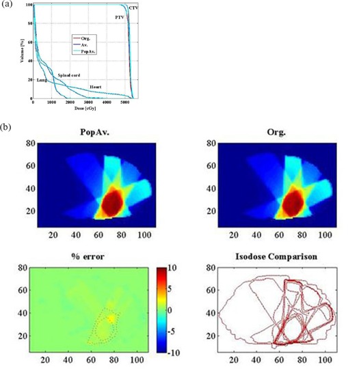
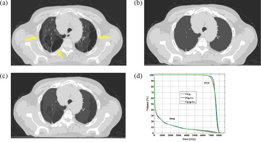
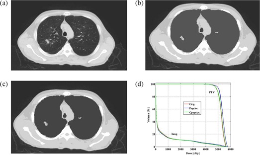
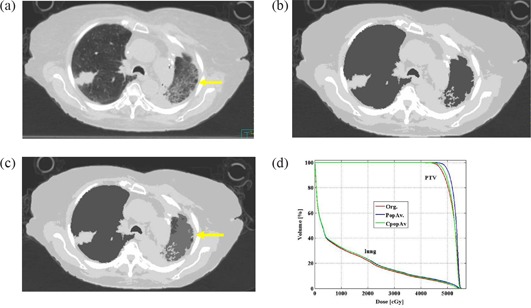
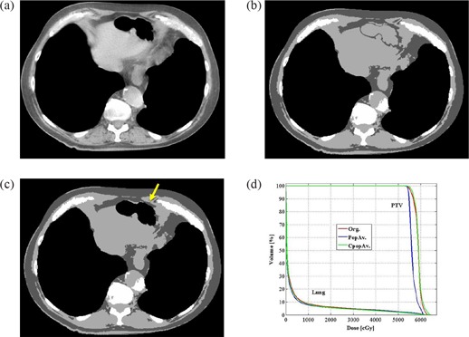
The aim of this study was to investigate the relative accuracy of megavoltage photon-beam dose calculations employing either 5 bulk densities or independent voxel densities determined by calibration of the CT Houndsfield number. Full-resolution CT and bulk density treatment plans were generated for 70 lung or esophageal cancer tumors (66 cases) using a commercial treatment planning system with an adaptive convolution dose calculation algorithm (Pinnacle3, Philips Medicals Systems). Bulk densities were applied to segmented regions. Individual and population average densities were compared to the full-resolution plan for each case. Monitor units were kept constant and no normalizations were employed. Dose volume histograms (DVH) and dose difference distributions were examined for all cases. The average densities of the segmented air, lung, fat, soft tissue, and bone for the entire set were found to be 0.14, 0.26, 0.89, 1.02, and 1.12 g/cc, respectively. In all cases, the normal tissue DVH agreed to better than 2% in dose. In 62 of 70 target DVHs, agreement to better than 3% in dose was observed. Six cases demonstrated emphysema, one with bullous formations and one with a hiatus hernia having a large volume of gas. They required the additional assignment of density to the emphysemic lung and inflammatory changes to the lung, the regions of collapsed lung, the bullous formations, and the hernia gas. Bulk tissue density dose calculation provides an accurate method of heterogeneous dose calculation. However, patients with advanced emphysema may require high-resolution CT studies for accurate treatment planning.
本研究的目的是调查使用5种整体密度或通过CT亨氏值校准确定的独立体素密度进行兆伏级光子束剂量计算的相对准确性。使用具有自适应卷积剂量计算算法的商业治疗计划系统(Pinnacle3,飞利浦医疗系统),为70例肺癌或食管癌肿瘤(66例患者)生成了全分辨率CT和整体密度治疗计划。将整体密度应用于分割区域。将每个病例的个体和总体平均密度与全分辨率计划进行比较。监测单位保持不变,未进行归一化处理。检查了所有病例的剂量体积直方图(DVH)和剂量差异分布。发现整个数据集的分割空气、肺、脂肪、软组织和骨骼的平均密度分别为0.14、0.26、0.89、1.02和1.12 g/cc。在所有病例中,正常组织的DVH在剂量方面的一致性优于2%。在70个靶区DVH中的62个中,观察到剂量一致性优于3%。6例显示肺气肿,1例有肺大泡形成,1例有食管裂孔疝且有大量气体。他们需要对肺气肿肺额外指定密度,并对肺、肺萎陷区域、肺大泡形成和疝气体区域的炎症变化指定密度。整体组织密度剂量计算提供了一种准确的非均匀剂量计算方法。然而,晚期肺气肿患者可能需要进行高分辨率CT研究以进行准确的治疗计划。