M310, Biomedical, Biomolecular and Chemical Sciences, Faculty of Life and Physical Sciences, The University of Western Australia, Western Australia, Australia.
Int Breastfeed J. 2009 Jun 1;4:5. doi: 10.1186/1746-4358-4-5.
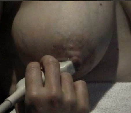
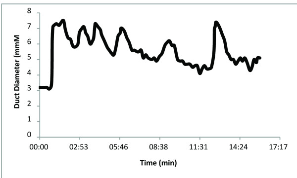
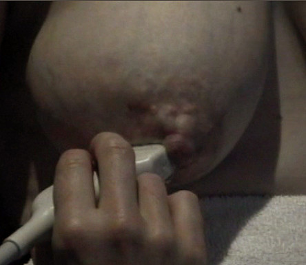
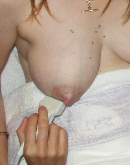
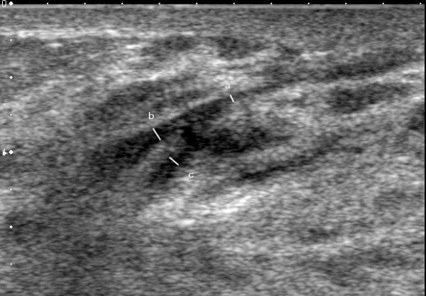
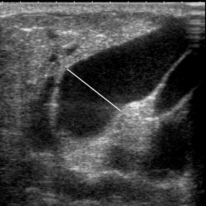
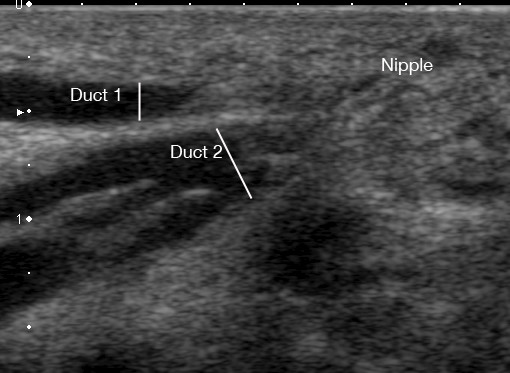
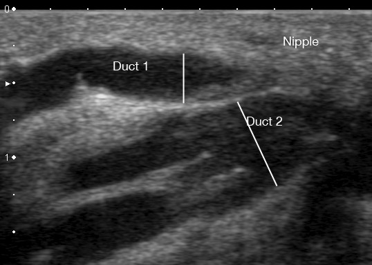
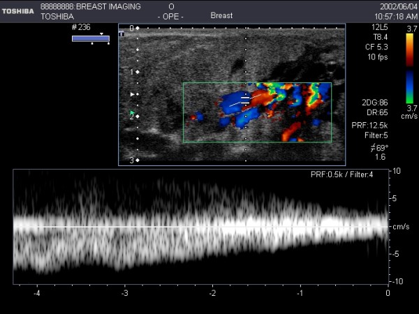
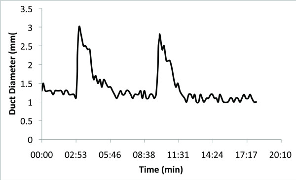
Diagnostic ultrasound imaging of the breast has been limited principally to the abnormal, non-lactating breast. Due to the rapid improvement of imaging technology, high-resolution ultrasound images can now be obtained of the lactating breast. Ultrasound scanning techniques, however, require modifications to accommodate the breast changes that occur in lactation. Furthermore, the function of the breast with regard to milk ejection can be assessed with ultrasound by identification of milk duct dilation and milk flow. At milk ejection, the echogenic duct walls expand as milk flows forward towards the nipple. Milk flow appears as echogenic foci rapidly moving within the milk duct. This paper provides a detailed description of the ultrasound technique used for the detection and reviews nuances associated with the procedure.
诊断性超声成像在乳腺方面的应用主要局限于异常的非哺乳期乳腺。由于成像技术的快速发展,现在可以获得高分辨率的哺乳期乳腺超声图像。然而,超声扫描技术需要进行修改,以适应哺乳期发生的乳房变化。此外,还可以通过识别乳腺导管扩张和乳汁流动来评估泌乳期乳房的功能。在泌乳期,随着乳汁向前流向乳头,高回声的导管壁扩张。乳汁流动表现为在乳腺导管内快速移动的高回声焦点。本文详细描述了用于检测的超声技术,并回顾了该过程的相关细微差别。