• 文献检索
  • 文档翻译
  • 深度研究
  • 学术资讯
  • Suppr Zotero 插件Zotero 插件
  • 邀请有礼
  • 套餐&价格
  • 历史记录
应用&插件
Suppr Zotero 插件Zotero 插件浏览器插件Mac 客户端Windows 客户端微信小程序
定价
高级版会员购买积分包购买API积分包
服务
文献检索文档翻译深度研究API 文档MCP 服务
关于我们
关于 Suppr公司介绍联系我们用户协议隐私条款
关注我们

Suppr 超能文献

核心技术专利:CN118964589B侵权必究
粤ICP备2023148730 号-1Suppr @ 2026

文献检索

告别复杂PubMed语法,用中文像聊天一样搜索,搜遍4000万医学文献。AI智能推荐,让科研检索更轻松。

立即免费搜索

文件翻译

保留排版,准确专业,支持PDF/Word/PPT等文件格式,支持 12+语言互译。

免费翻译文档

深度研究

AI帮你快速写综述,25分钟生成高质量综述,智能提取关键信息,辅助科研写作。

立即免费体验

主动脉瓣疾病中左心室肥厚的心电图诊断:通过心血管磁共振评估心电图标准

Electrocardiographic diagnosis of left ventricular hypertrophy in aortic valve disease: evaluation of ECG criteria by cardiovascular magnetic resonance.

作者信息

Buchner Stefan, Debl Kurt, Haimerl Josef, Djavidani Behrus, Poschenrieder Florian, Feuerbach Stefan, Riegger Guenter A J, Luchner Andreas

机构信息

Klinik und Poliklinik für Innere Medizin II, Universitätsklinikum Regensburg, Germany.

出版信息

J Cardiovasc Magn Reson. 2009 Jun 1;11(1):18. doi: 10.1186/1532-429X-11-18.

DOI:10.1186/1532-429X-11-18
PMID:19486532
原文链接:https://pmc.ncbi.nlm.nih.gov/articles/PMC2696426/
Abstract

BACKGROUND

Left ventricular hypertrophy (LVH) is a hallmark of chronic pressure or volume overload of the left ventricle and is associated with risk of cardiovascular morbidity and mortality. The purpose was to evaluate different electrocardiographic criteria for LVH as determined by cardiovascular magnetic resonance (CMR). Additionally, the effects of concentric and eccentric LVH on depolarization and repolarization were assessed.

METHODS

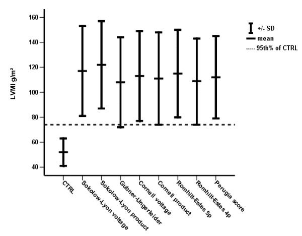
120 patients with aortic valve disease and 30 healthy volunteers were analysed. As ECG criteria for LVH, we assessed the Sokolow-Lyon voltage/product, Gubner-Ungerleider voltage, Cornell voltage/product, Perugia-score and Romhilt-Estes score.

RESULTS

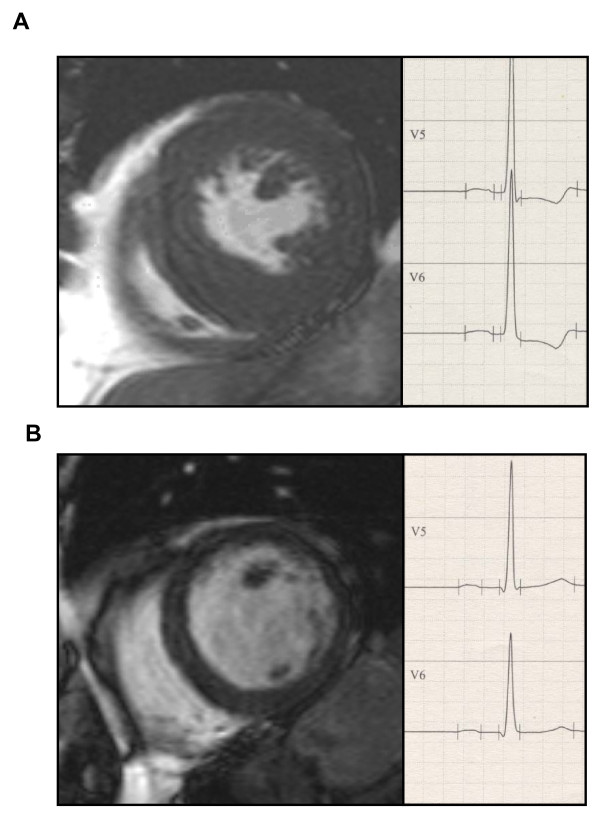
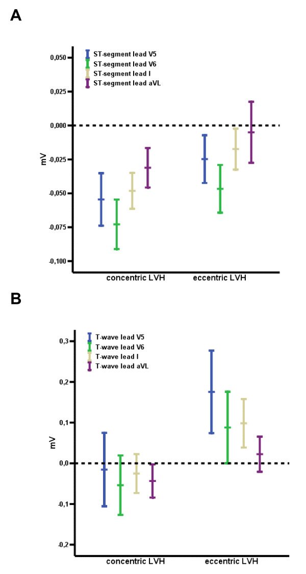
All ECG criteria demonstrated a significant correlation with LV mass and chamber size. The highest predictive values were achieved by the Romhilt-Estes score 4 points with a sensitivity of 86% and specificity of 81%. There was no difference in all ECG criteria between concentric and eccentric LVH. However, the intrinsicoid deflection (V6 37 +/- 1.0 ms vs. 43 +/- 1.6 ms, p < 0.05) was shorter in concentric LVH than in eccentric LVH and amplitudes of ST-segment (V5 -0.06 +/- 0.01 vs. -0.02 +/- 0.01) and T-wave (V5 -0.03 +/- 0.04 vs. 0.18 +/- 0.05) in the anterolateral leads (p < 0.05) were deeper.

CONCLUSION

By calibration with CMR, a wide range of predictive values was found for the various ECG criteria for LVH with the most favourable results for the Romhilt-Estes score. As electrocardiographic correlate for concentric LVH as compared with eccentric LVH, a shorter intrinsicoid deflection and a significant ST-segment and T-wave depression in the anterolateral leads was noted.

摘要

背景

左心室肥厚(LVH)是左心室慢性压力或容量超负荷的标志,与心血管疾病的发病率和死亡率风险相关。目的是评估心血管磁共振(CMR)测定的不同LVH心电图标准。此外,还评估了向心性和离心性LVH对去极化和复极化的影响。

方法

分析了120例主动脉瓣疾病患者和30名健康志愿者。作为LVH的心电图标准,我们评估了索科洛 - 里昂电压/乘积、古布纳 - 昂格勒德电压、康奈尔电压/乘积、佩鲁贾评分和罗米尔 - 埃斯评分。

结果

所有心电图标准均与左心室质量和腔室大小显著相关。罗米尔 - 埃斯评分4分时预测价值最高,敏感性为86%,特异性为81%。向心性和离心性LVH在所有心电图标准上无差异。然而,向心性LVH的类本位曲折(V6 37±1.0毫秒对43±1.6毫秒,p<0.05)比离心性LVH短,前外侧导联的ST段振幅(V5 -0.06±0.01对-0.02±0.01)和T波振幅(V5 -0.03±0.04对0.18±0.05)更深(p<0.05)。

结论

通过与CMR校准,发现各种LVH心电图标准具有广泛的预测价值,罗米尔 - 埃斯评分结果最佳。与离心性LVH相比,向心性LVH的心电图相关性表现为类本位曲折较短,前外侧导联有明显的ST段和T波压低。

https://cdn.ncbi.nlm.nih.gov/pmc/blobs/170b/2696426/dda942ce98f5/1532-429X-11-18-4.jpg
https://cdn.ncbi.nlm.nih.gov/pmc/blobs/170b/2696426/3e5e57c94a23/1532-429X-11-18-1.jpg
https://cdn.ncbi.nlm.nih.gov/pmc/blobs/170b/2696426/e423f3588a84/1532-429X-11-18-2.jpg
https://cdn.ncbi.nlm.nih.gov/pmc/blobs/170b/2696426/b36dbe9e3079/1532-429X-11-18-3.jpg
https://cdn.ncbi.nlm.nih.gov/pmc/blobs/170b/2696426/dda942ce98f5/1532-429X-11-18-4.jpg

相似文献

1
Electrocardiographic diagnosis of left ventricular hypertrophy in aortic valve disease: evaluation of ECG criteria by cardiovascular magnetic resonance.主动脉瓣疾病中左心室肥厚的心电图诊断:通过心血管磁共振评估心电图标准
J Cardiovasc Magn Reson. 2009 Jun 1;11(1):18. doi: 10.1186/1532-429X-11-18.
2
The effect of obesity on electrocardiographic detection of hypertensive left ventricular hypertrophy: recalibration against cardiac magnetic resonance.肥胖对高血压性左心室肥厚心电图检测的影响:与心脏磁共振成像对比重新校准
J Hum Hypertens. 2016 Mar;30(3):197-203. doi: 10.1038/jhh.2015.58. Epub 2015 Jun 4.
3
Standard electrocardiographic criteria for left ventricular hypertrophy in Nigerian hypertensives.尼日利亚高血压患者左心室肥厚的标准心电图诊断标准
Ethn Dis. 2005 Autumn;15(4):578-84.
4
Accuracy of ECG criteria for the diagnosis of left ventricular hypertrophy: a comparison with magnetic resonance imaging.心电图诊断左心室肥厚标准的准确性:与磁共振成像的比较
J Med Assoc Thai. 2013 Feb;96 Suppl 2:S124-32.
5
The relationship between electrocardiographic left ventricular hypertrophy criteria and echocardiographic mass in patients undergoing transcatheter aortic valve replacement.经导管主动脉瓣置换术患者心电图左心室肥厚标准与超声心动图测量的左心室质量之间的关系。
J Electrocardiol. 2015 Jul-Aug;48(4):630-6. doi: 10.1016/j.jelectrocard.2015.03.008. Epub 2015 Mar 11.
6
Comparison of Araoye's criteria with standard electrocardiographic criteria for diagnosis of left ventricular hypertrophy in Nigerian hypertensives.在尼日利亚高血压患者中,Araoye标准与诊断左心室肥厚的标准心电图标准的比较。
West Afr J Med. 2006 Jul-Sep;25(3):179-85. doi: 10.4314/wajm.v25i3.28274.
7
Diagnostic performance of electrocardiographic criteria for left ventricular hypertrophy among various body mass index groups compared to diagnosis by cardiac magnetic resonance imaging.与心脏磁共振成像诊断相比,不同体重指数组中左心室肥厚心电图标准的诊断性能。
Ann Noninvasive Electrocardiol. 2019 Jul;24(4):e12635. doi: 10.1111/anec.12635. Epub 2019 Feb 5.
8
Evaluation of ECG criteria for left ventricular hypertrophy before and after aortic valve replacement using magnetic resonance imaging.使用磁共振成像评估主动脉瓣置换术前和术后左心室肥厚的心电图标准
J Cardiovasc Magn Reson. 2003 Jul;5(3):465-74. doi: 10.1081/jcmr-120022262.
9
Prognostic value of a new electrocardiographic method for diagnosis of left ventricular hypertrophy in essential hypertension.一种用于诊断原发性高血压左心室肥厚的新心电图方法的预后价值
J Am Coll Cardiol. 1998 Feb;31(2):383-90. doi: 10.1016/s0735-1097(97)00493-2.
10
Evaluation of the electrocardiographic criteria for left ventricular hypertrophy.左心室肥厚心电图标准的评估。
Anadolu Kardiyol Derg. 2007 Jul;7 Suppl 1:159-63.

引用本文的文献

1
Electrocardiographic R Wave Peak Time: Correlation With the Severity of Coronary Artery Disease and Prognosis in Patients With Acute Coronary Syndrome.心电图R波峰时间:与急性冠状动脉综合征患者冠状动脉疾病严重程度及预后的相关性
Catheter Cardiovasc Interv. 2025 Aug;106(2):1225-1232. doi: 10.1002/ccd.31669. Epub 2025 Jun 8.
2
Cardiac Morphology, Function, and Left Ventricular Geometric Pattern in Patients with Hypertensive Crisis: A Cardiovascular Magnetic Resonance-Based Study.高血压急症患者的心脏形态、功能及左心室几何形态模式:一项基于心血管磁共振成像的研究
J Cardiovasc Dev Dis. 2023 Aug 27;10(9):367. doi: 10.3390/jcdd10090367.
3

本文引用的文献

1
The ventricular complex in left ventricular hypertrophy as obtained by unipolar precordial and limb leads.通过单极胸前导联和肢体导联获得的左心室肥厚时的心室复合波。
Am Heart J. 1949 Feb;37(2):161-86. doi: 10.1016/0002-8703(49)90562-1.
2
Cardiovascular function in multi-ethnic study of atherosclerosis: normal values by age, sex, and ethnicity.动脉粥样硬化多民族研究中的心血管功能:按年龄、性别和种族划分的正常值
AJR Am J Roentgenol. 2006 Jun;186(6 Suppl 2):S357-65. doi: 10.2214/AJR.04.1868.
3
Association between electrocardiographic left ventricular hypertrophy with strain pattern and left ventricular structure and function.
Clinical Data, Chest Radiograph and Electrocardiography in the Screening for Left Ventricular Hypertrophy: The CARE Score.
用于筛查左心室肥厚的临床数据、胸部X光片和心电图:CARE评分
J Clin Med. 2022 Jun 22;11(13):3585. doi: 10.3390/jcm11133585.
4
Implication of Different ECG Left Ventricular Hypertrophy in Patients Undergoing Transcatheter Aortic Valve Replacement.不同心电图左心室肥厚在经导管主动脉瓣置换术患者中的意义。
J Am Heart Assoc. 2022 Feb 15;11(4):e023647. doi: 10.1161/JAHA.121.023647. Epub 2022 Feb 3.
5
Electrocardiographic criteria for left ventricular hypertrophy in aortic valve stenosis: Correlation with echocardiographic parameters.主动脉瓣狭窄时左心室肥厚的心电图标准:与超声心动图参数的相关性。
Ann Noninvasive Electrocardiol. 2019 Sep;24(5):e12645. doi: 10.1111/anec.12645. Epub 2019 Mar 21.
6
Detection of Left Ventricular Hypertrophy Using Bayesian Additive Regression Trees: The MESA.贝叶斯加法回归树检测左心室肥厚:MESA。
J Am Heart Assoc. 2019 Mar 5;8(5):e009959. doi: 10.1161/JAHA.118.009959.
7
Diagnostic performance of electrocardiographic criteria for left ventricular hypertrophy among various body mass index groups compared to diagnosis by cardiac magnetic resonance imaging.与心脏磁共振成像诊断相比,不同体重指数组中左心室肥厚心电图标准的诊断性能。
Ann Noninvasive Electrocardiol. 2019 Jul;24(4):e12635. doi: 10.1111/anec.12635. Epub 2019 Feb 5.
8
Electrocardiographic left ventricular hypertrophy predicts recurrence of atrial arrhythmias after catheter ablation of paroxysmal atrial fibrillation.心电图左心室肥厚可预测阵发性心房颤动导管消融术后房性心律失常的复发。
Clin Cardiol. 2018 Jun;41(6):797-802. doi: 10.1002/clc.22957. Epub 2018 Jun 5.
9
Noctural dipping status and left ventricular hypertrophy: A cardiac magnetic resonance imaging study.夜间血压下降情况与左心室肥厚:一项心脏磁共振成像研究。
J Clin Hypertens (Greenwich). 2018 Apr;20(4):784-793. doi: 10.1111/jch.13235. Epub 2018 Mar 8.
10
Electrocardiographic Strain Pattern Is Associated With Left Ventricular Concentric Remodeling, Scar, and Mortality Over 10 Years: The Multi-Ethnic Study of Atherosclerosis.心电图应变模式与左心室向心性重构、瘢痕和 10 年后的死亡率相关:动脉粥样硬化多民族研究。
J Am Heart Assoc. 2017 Sep 20;6(9):e006624. doi: 10.1161/JAHA.117.006624.
伴有应变模式的心电图左心室肥厚与左心室结构和功能之间的关联
Cardiology. 2006;106(1):14-21. doi: 10.1159/000092478. Epub 2006 Apr 4.
4
Controversies in ventricular remodelling.心室重构中的争议
Lancet. 2006 Jan 28;367(9507):356-67. doi: 10.1016/S0140-6736(06)68074-4.
5
Normal human left and right ventricular and left atrial dimensions using steady state free precession magnetic resonance imaging.使用稳态自由进动磁共振成像测量正常人体左、右心室及左心房尺寸。
J Cardiovasc Magn Reson. 2005;7(5):775-82. doi: 10.1080/10976640500295516.
6
Planimetry of aortic valve area in aortic stenosis by magnetic resonance imaging.磁共振成像评估主动脉瓣狭窄时主动脉瓣面积的平面测量法
Invest Radiol. 2005 Oct;40(10):631-6. doi: 10.1097/01.rli.0000178362.67085.fd.
7
Quantification of aortic valve area and left ventricular muscle mass in healthy subjects and patients with symptomatic aortic valve stenosis by MRI.通过磁共振成像(MRI)对健康受试者和有症状主动脉瓣狭窄患者的主动脉瓣面积和左心室肌肉质量进行量化。
Z Kardiol. 2005 Mar;94(3):173-81. doi: 10.1007/s00392-005-0198-1.
8
Early regression of left ventricular hypertrophy after aortic valve replacement by the Ross procedure detected by cine MRI.通过电影磁共振成像检测到Ross手术置换主动脉瓣后左心室肥厚的早期消退。
J Cardiovasc Magn Reson. 2004;6(1):1-8. doi: 10.1081/jcmr-120027799.
9
Systolic and diastolic loading of the heart. I. Physiologic and clinical data.
Am Heart J. 1952 May;43(5):661-8. doi: 10.1016/0002-8703(52)90041-0.
10
Normal human left and right ventricular dimensions for MRI as assessed by turbo gradient echo and steady-state free precession imaging sequences.通过涡轮梯度回波和稳态自由进动成像序列评估的正常人类心脏磁共振成像左右心室尺寸。
J Magn Reson Imaging. 2003 Mar;17(3):323-9. doi: 10.1002/jmri.10262.