Djaberi Roxana, Schuijf Joanne D, Boersma Eric, Kroft Lucia J M, Pereira Alberto M, Romijn Johannes A, Scholte Arthur J, Jukema J Wouter, Bax Jeroen J
Department of Cardiology, Leiden University Medical Center, Leiden, the Netherlands.
Diabetes Care. 2009 Aug;32(8):1507-12. doi: 10.2337/dc09-0320. Epub 2009 Jun 1.
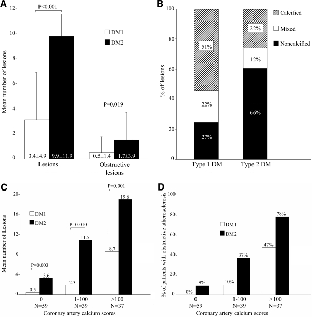
OBJECTIVE It is unclear whether the coronary atherosclerotic plaque burden is similar in patients with type 1 and type 2 diabetes. By using multislice computed tomography (MSCT), the presence, degree, and morphology of coronary artery disease (CAD) in patients with type 1 and type 2 diabetes were compared. RESEARCH DESIGN AND METHODS Prospectively, coronary artery calcium (CAC) scoring and MSCT coronary angiography were performed in 135 asymptomatic patients (65 patients with type 1 diabetes and 70 patients with type 2 diabetes). The presence and extent of coronary atherosclerosis as well as plaque phenotype were assessed and compared between groups. RESULTS No difference was observed in average CAC score (217 +/- 530 vs. 174 +/- 361) or in the prevalence of coronary atherosclerosis (65% vs. 71%) in patients with type 1 and type 2 diabetes. However, the prevalence of obstructive atherosclerosis was higher in patients with type 2 diabetes (n = 24; 34%) compared with that in patients with type 1 diabetes (n = 11; 17%) (P = 0.02). In addition, a higher mean number of atherosclerotic and obstructive plaques was observed in patients with type 2 diabetes. In addition, the percentage of noncalcified plaques was higher in patients with type 2 (66%) versus type 1 diabetes (27%) (P < 0.001), resulting in a higher plaque burden for each CAC score compared with that in type 1 diabetic patients. CONCLUSIONS Although CAC scores and the prevalence of coronary atherosclerosis were similar between patients with type 1 and type 2 diabetes, CAD was more extensive in the latter. Also, a relatively higher proportion of noncalcified plaques was observed in patients with type 2 diabetes. These observations may be valuable in the development of targeted management strategies adapted to diabetes type.
目的 1 型糖尿病患者和 2 型糖尿病患者的冠状动脉粥样硬化斑块负荷是否相似尚不清楚。通过使用多层螺旋计算机断层扫描(MSCT),比较了 1 型糖尿病患者和 2 型糖尿病患者冠状动脉疾病(CAD)的存在、程度和形态。
研究设计与方法 前瞻性地对 135 例无症状患者(65 例 1 型糖尿病患者和 70 例 2 型糖尿病患者)进行冠状动脉钙化(CAC)评分和 MSCT 冠状动脉造影。评估并比较两组之间冠状动脉粥样硬化的存在和程度以及斑块表型。
结果 1 型糖尿病患者和 2 型糖尿病患者的平均 CAC 评分(217±530 与 174±361)或冠状动脉粥样硬化患病率(65%与 71%)无差异。然而,2 型糖尿病患者的阻塞性动脉粥样硬化患病率(n = 24;34%)高于 1 型糖尿病患者(n = 11;17%)(P = 0.02)。此外,2 型糖尿病患者观察到的动脉粥样硬化斑块和阻塞性斑块的平均数量更高。此外,2 型糖尿病患者(66%)的非钙化斑块百分比高于 1 型糖尿病患者(27%)(P < 0.001),与 1 型糖尿病患者相比,每个 CAC 评分的斑块负荷更高。
结论 尽管 1 型糖尿病患者和 2 型糖尿病患者的 CAC 评分和冠状动脉粥样硬化患病率相似,但后者的 CAD 更为广泛。此外,在 2 型糖尿病患者中观察到相对较高比例的非钙化斑块。这些观察结果可能对制定适合糖尿病类型的靶向管理策略具有重要价值。