Kobrinsky Evgeny, Abrahimi Parwiz, Duong Son Q, Thomas Sam, Harry Jo Beth, Patel Chirag, Lao Qi Zong, Soldatov Nikolai M
National Institute on Aging, National Institutes of Health, Baltimore, Maryland, United States of America.
PLoS One. 2009 May 18;4(5):e5587. doi: 10.1371/journal.pone.0005587.
Voltage-gated Ca(v)1.2 calcium channels play a crucial role in Ca(2+) signaling. The pore-forming alpha(1C) subunit is regulated by accessory Ca(v)beta subunits, cytoplasmic proteins of various size encoded by four different genes (Ca(v)beta(1)-beta(4)) and expressed in a tissue-specific manner.
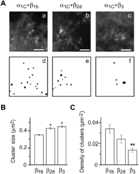
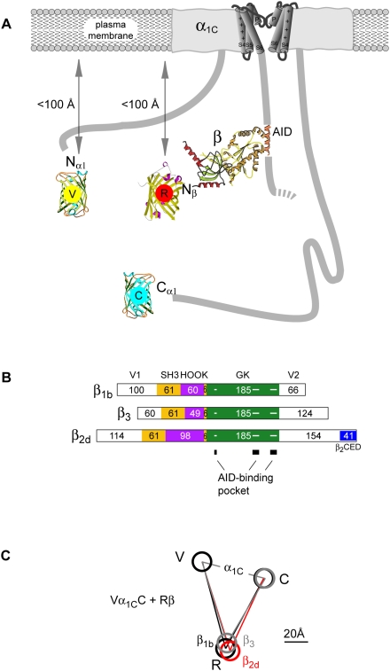
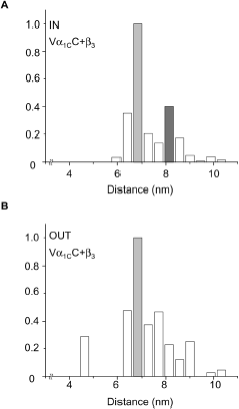
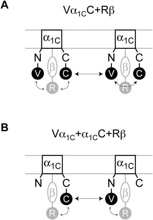
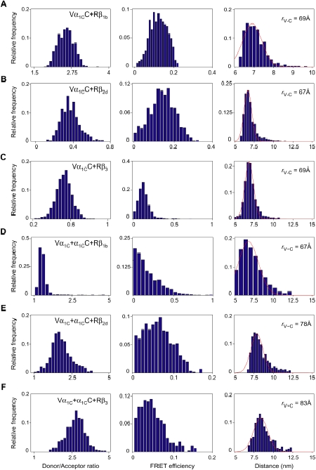
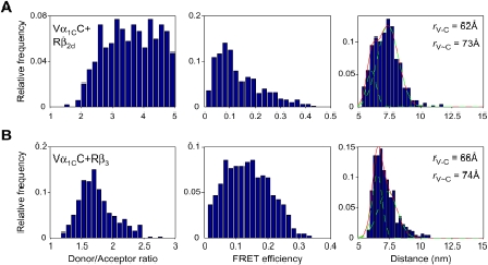
Here we investigated the effect of three major Ca(v)beta types, beta(1b), beta(2d) and beta(3), on the structure of Ca(v)1.2 in the plasma membrane of live cells. Total internal reflection fluorescence microscopy showed that the tendency of Ca(v)1.2 to form clusters depends on the type of the Ca(v)beta subunit present. The highest density of Ca(v)1.2 clusters in the plasma membrane and the smallest cluster size were observed with neuronal/cardiac beta(1b) present. Ca(v)1.2 channels containing beta(3), the predominant Ca(v)beta subunit of vascular smooth muscle cells, were organized in a significantly smaller number of larger clusters. The inter- and intramolecular distances between alpha(1C) and Ca(v)beta in the plasma membrane of live cells were measured by three-color FRET microscopy. The results confirm that the proximity of Ca(v)1.2 channels in the plasma membrane depends on the Ca(v)beta type. The presence of different Ca(v)beta subunits does not result in significant differences in the intramolecular distance between the termini of alpha(1C), but significantly affects the distance between the termini of neighbor alpha(1C) subunits, which varies from 67 A with beta(1b) to 79 A with beta(3).
Thus, our results show that the structural organization of Ca(v)1.2 channels in the plasma membrane depends on the type of Ca(v)beta subunits present.
电压门控性Ca(v)1.2钙通道在Ca(2+)信号传导中起关键作用。形成孔道的α(1C)亚基受辅助性Ca(v)β亚基调节,Ca(v)β亚基是由四个不同基因(Ca(v)β(1)-β(4))编码的各种大小的胞质蛋白,并以组织特异性方式表达。
在此,我们研究了三种主要的Ca(v)β类型,即β(1b)、β(2d)和β(3),对活细胞质膜中Ca(v)1.2结构的影响。全内反射荧光显微镜显示,Ca(v)1.2形成簇的倾向取决于所存在的Ca(v)β亚基的类型。当存在神经元/心脏型β(1b)时,观察到质膜中Ca(v)1.2簇的密度最高且簇的尺寸最小。含有β(3)(血管平滑肌细胞的主要Ca(v)β亚基)的Ca(v)1.2通道以明显更少数量的更大簇的形式组织。通过三色荧光共振能量转移显微镜测量活细胞质膜中α(1C)和Ca(v)β之间的分子间和分子内距离。结果证实,质膜中Ca(v)1.2通道的接近程度取决于Ca(v)β类型。不同Ca(v)β亚基的存在不会导致α(1C)末端之间分子内距离的显著差异,但会显著影响相邻α(1C)亚基末端之间的距离,该距离从与β(1b)结合时的67 Å变化到与β(3)结合时的79 Å。
因此,我们的结果表明,质膜中Ca(v)1.2通道的结构组织取决于所存在的Ca(v)β亚基的类型。