• 文献检索
  • 文档翻译
  • 深度研究
  • 学术资讯
  • Suppr Zotero 插件Zotero 插件
  • 邀请有礼
  • 套餐&价格
  • 历史记录
应用&插件
Suppr Zotero 插件Zotero 插件浏览器插件Mac 客户端Windows 客户端微信小程序
定价
高级版会员购买积分包购买API积分包
服务
文献检索文档翻译深度研究API 文档MCP 服务
关于我们
关于 Suppr公司介绍联系我们用户协议隐私条款
关注我们

Suppr 超能文献

核心技术专利:CN118964589B侵权必究
粤ICP备2023148730 号-1Suppr @ 2026

文献检索

告别复杂PubMed语法,用中文像聊天一样搜索,搜遍4000万医学文献。AI智能推荐,让科研检索更轻松。

立即免费搜索

文件翻译

保留排版,准确专业,支持PDF/Word/PPT等文件格式,支持 12+语言互译。

免费翻译文档

深度研究

AI帮你快速写综述,25分钟生成高质量综述,智能提取关键信息,辅助科研写作。

立即免费体验

颅内动脉瘤性蛛网膜下腔出血后血管痉挛、脑灌注与迟发性脑缺血的关系。

Relationship between vasospasm, cerebral perfusion, and delayed cerebral ischemia after aneurysmal subarachnoid hemorrhage.

机构信息

Department of Radiology (HP E01.132), University Medical Center, P. O. Box 85500, 3508 GA, Utrecht, The Netherlands.

出版信息

Neuroradiology. 2009 Dec;51(12):813-9. doi: 10.1007/s00234-009-0575-y. Epub 2009 Jul 22.

DOI:10.1007/s00234-009-0575-y
PMID:19623472
原文链接:https://pmc.ncbi.nlm.nih.gov/articles/PMC2773037/
Abstract

INTRODUCTION

Vasospasm after aneurysmal subarachnoid hemorrhage (SAH) is thought to cause ischemia. To evaluate the contribution of vasospasm to delayed cerebral ischemia (DCI), we investigated the effect of vasospasm on cerebral perfusion and the relationship of vasospasm with DCI.

METHODS

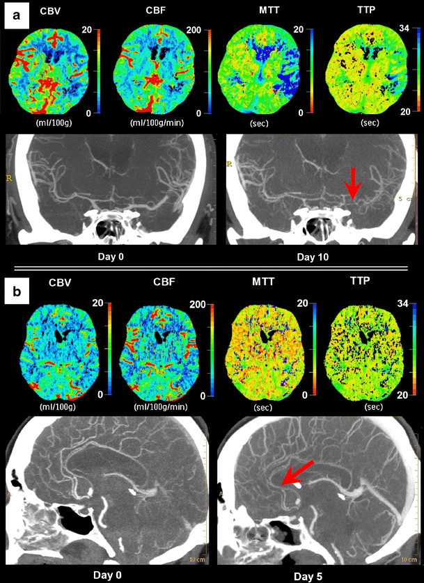
We studied 37 consecutive SAH patients with CT angiography (CTA) and CT perfusion (CTP) on admission and within 14 days after admission or at time of clinical deterioration. CTP values (cerebral blood volume, cerebral blood flow (CBF) and mean transit time), degree of vasospasm on CTA, and occurrence of DCI were recorded. Vasospasm was categorized as follows: no spasm (0-25% decrease in vessel diameter), moderate spasm (25-50% decrease), and severe spasm (>50% decrease). The correspondence of the flow territory of the most spastic vessel with the least perfused region was evaluated, and differences in perfusion values and occurrence of DCI between degrees of vasospasm were calculated with 95% confidence intervals (95% CI).

RESULTS

Fourteen patients had no vasospasm, 16 were moderate, and seven were severe. In 65% of patients with spasm, the flow territory of the most spastic vessel corresponded with the least perfused region. There was significant CBF (milliliters per 100 g per minute) difference (-21.3; 95% CI, -37 <--> -5.3) between flow territories of severe and no vasospasm. Four of seven patients with severe, six of 16 with moderate, and three of 14 patients with no vasospasm had DCI.

CONCLUSION

Vasospasm decreases cerebral perfusion, but corresponds with the least perfused region in only two thirds of our patients. Furthermore, almost half of patients with severe vasospasm do not have DCI. Thus, although severe vasospasm can decrease perfusion, it may not result in DCI.

摘要

介绍

人们认为,蛛网膜下腔出血(SAH)后的血管痉挛会导致缺血。为了评估血管痉挛对迟发性脑缺血(DCI)的影响,我们研究了血管痉挛对脑灌注的影响,以及血管痉挛与 DCI 的关系。

方法

我们研究了 37 例连续的 SAH 患者,他们在入院时和入院后 14 天内或临床恶化时进行了 CT 血管造影(CTA)和 CT 灌注(CTP)检查。记录了 CTP 值(脑血容量、脑血流(CBF)和平均通过时间)、CTA 上的血管痉挛程度以及 DCI 的发生情况。血管痉挛分为:无痉挛(血管直径减少 0-25%)、中度痉挛(血管直径减少 25-50%)和重度痉挛(血管直径减少>50%)。评估最痉挛血管的血流区域与最灌注不足区域的对应关系,并计算不同程度血管痉挛之间的灌注值和 DCI 的发生率差异,置信区间为 95%(95%CI)。

结果

14 例患者无血管痉挛,16 例为中度痉挛,7 例为重度痉挛。在 65%的痉挛患者中,最痉挛血管的血流区域与最灌注不足区域相对应。严重和无痉挛之间的 CBF(每分钟每 100 克毫升)差异有统计学意义(-21.3;95%CI,-37 <--> -5.3)。7 例重度痉挛患者中有 4 例、16 例中度痉挛患者中有 6 例、14 例无痉挛患者中有 3 例发生 DCI。

结论

血管痉挛会降低脑灌注,但在我们的患者中只有三分之二的情况下与最灌注不足的区域相对应。此外,几乎一半的重度血管痉挛患者没有发生 DCI。因此,尽管重度血管痉挛会降低灌注,但它可能不会导致 DCI。

https://cdn.ncbi.nlm.nih.gov/pmc/blobs/5d50/2773037/b3317da867e7/234_2009_575_Fig2_HTML.jpg
https://cdn.ncbi.nlm.nih.gov/pmc/blobs/5d50/2773037/35a4539f57f8/234_2009_575_Fig1_HTML.jpg
https://cdn.ncbi.nlm.nih.gov/pmc/blobs/5d50/2773037/b3317da867e7/234_2009_575_Fig2_HTML.jpg
https://cdn.ncbi.nlm.nih.gov/pmc/blobs/5d50/2773037/35a4539f57f8/234_2009_575_Fig1_HTML.jpg
https://cdn.ncbi.nlm.nih.gov/pmc/blobs/5d50/2773037/b3317da867e7/234_2009_575_Fig2_HTML.jpg

相似文献

1
Relationship between vasospasm, cerebral perfusion, and delayed cerebral ischemia after aneurysmal subarachnoid hemorrhage.颅内动脉瘤性蛛网膜下腔出血后血管痉挛、脑灌注与迟发性脑缺血的关系。
Neuroradiology. 2009 Dec;51(12):813-9. doi: 10.1007/s00234-009-0575-y. Epub 2009 Jul 22.
2
Cost-effectiveness of CT angiography and perfusion imaging for delayed cerebral ischemia and vasospasm in aneurysmal subarachnoid hemorrhage.CT血管造影和灌注成像对动脉瘤性蛛网膜下腔出血后迟发性脑缺血和血管痉挛的成本效益分析
AJNR Am J Neuroradiol. 2014 Sep;35(9):1714-20. doi: 10.3174/ajnr.A3947. Epub 2014 May 8.
3
Early low cerebral blood flow and high cerebral lactate: prediction of delayed cerebral ischemia in subarachnoid hemorrhage.早期脑血流低和脑乳酸高:蛛网膜下腔出血后迟发性脑缺血的预测。
J Neurosurg. 2018 Jun;128(6):1762-1770. doi: 10.3171/2016.11.JNS161140. Epub 2017 Jun 2.
4
Early Perfusion Computed Tomography Scan for Prediction of Vasospasm and Delayed Cerebral Ischemia After Aneurysmal Subarachnoid Hemorrhage.早期灌注 CT 扫描预测动脉瘤性蛛网膜下腔出血后血管痉挛和迟发性脑缺血。
World Neurosurg. 2019 Oct;130:e743-e752. doi: 10.1016/j.wneu.2019.06.213. Epub 2019 Jul 5.
5
Whole brain CT perfusion combined with CT angiography in patients with subarachnoid hemorrhage and cerebral vasospasm.全脑CT灌注联合CT血管造影在蛛网膜下腔出血和脑血管痉挛患者中的应用
Clin Neurol Neurosurg. 2013 Dec;115(12):2496-501. doi: 10.1016/j.clineuro.2013.10.004. Epub 2013 Oct 16.
6
Advancing diagnostic precision of delayed cerebral ischemia in aneurysmal subarachnoid hemorrhage: The potential for a vasospasm index score on perfusion imaging to detect vasospasm.提高动脉瘤性蛛网膜下腔出血后迟发性脑缺血的诊断精度:灌注成像血管痉挛指数评分检测血管痉挛的潜力。
Eur J Radiol. 2024 Sep;178:111578. doi: 10.1016/j.ejrad.2024.111578. Epub 2024 Jun 27.
7
Early whole-brain CT perfusion for detection of patients at risk for delayed cerebral ischemia after subarachnoid hemorrhage.早期全脑 CT 灌注检测蛛网膜下腔出血后迟发性脑缺血高危患者。
J Neurosurg. 2016 Jul;125(1):128-36. doi: 10.3171/2015.6.JNS15720. Epub 2015 Dec 18.
8
Disturbance of CT perfusion within 24 h after onset is associated with WFNS grade but not development of DCI in patients with aneurysmal SAH.动脉瘤性蛛网膜下腔出血患者发病后24小时内的CT灌注紊乱与WFNS分级相关,但与迟发性脑缺血的发生无关。
Acta Neurochir (Wien). 2017 Dec;159(12):2319-2324. doi: 10.1007/s00701-017-3362-2. Epub 2017 Oct 23.
9
CT angiography and perfusion imaging in patients with subarachnoid hemorrhage: correlation of vasospasm to perfusion abnormality.蛛网膜下腔出血患者的CT血管造影和灌注成像:血管痉挛与灌注异常的相关性
Neuroradiology. 2009 Feb;51(2):85-93. doi: 10.1007/s00234-008-0466-7. Epub 2008 Oct 11.
10
Perfusion Computed Tomography as a Screening Tool for Pending Delayed Cerebral Ischemia in Comatose Patients After Aneurysmal Subarachnoid Hemorrhage: A Retrospective Cohort Study.灌注计算机断层扫描作为动脉瘤性蛛网膜下腔出血后昏迷患者迟发性脑缺血的筛查工具:一项回顾性队列研究。
Neurocrit Care. 2024 Jun;40(3):964-975. doi: 10.1007/s12028-023-01855-6. Epub 2023 Oct 11.

引用本文的文献

1
Cerebral vasospasm following aneurysmal subarachnoid hemorrhage: the impact of cocaine use, Hunt-Hess grade, and other risk factors.动脉瘤性蛛网膜下腔出血后的脑血管痉挛:可卡因使用、Hunt-Hess分级及其他危险因素的影响
Neuroradiology. 2025 Jul 28. doi: 10.1007/s00234-025-03713-y.
2
Effects of metformin use on aneurysmal subarachnoid hemorrhage outcomes.二甲双胍的使用对动脉瘤性蛛网膜下腔出血预后的影响。
Acta Neurochir (Wien). 2025 Apr 10;167(1):103. doi: 10.1007/s00701-025-06516-5.
3
The clinical impact of recent amphetamine exposure in aneurysmal subarachnoid hemorrhage patients.

本文引用的文献

1
CT angiography and perfusion imaging in patients with subarachnoid hemorrhage: correlation of vasospasm to perfusion abnormality.蛛网膜下腔出血患者的CT血管造影和灌注成像:血管痉挛与灌注异常的相关性
Neuroradiology. 2009 Feb;51(2):85-93. doi: 10.1007/s00234-008-0466-7. Epub 2008 Oct 11.
2
Perfusion-weighted MRI to evaluate cerebral autoregulation in aneurysmal subarachnoid haemorrhage.灌注加权磁共振成像评估动脉瘤性蛛网膜下腔出血的脑自动调节功能
Neuroradiology. 2008 Nov;50(11):929-38. doi: 10.1007/s00234-008-0424-4. Epub 2008 Jun 17.
3
Visual grading system for vasospasm based on perfusion CT imaging: comparisons with conventional angiography and quantitative perfusion CT.
近期使用苯丙胺对动脉瘤性蛛网膜下腔出血患者的临床影响。
Front Neurol. 2025 Jan 7;15:1480401. doi: 10.3389/fneur.2024.1480401. eCollection 2024.
4
Cilostazol Alleviates Delayed Cerebral Ischemia After Subarachnoid Hemorrhage by Attenuating Microcirculatory Dysfunction.西洛他唑通过减轻微循环功能障碍减轻蛛网膜下腔出血后的迟发性脑缺血。
Transl Stroke Res. 2024 Nov 20. doi: 10.1007/s12975-024-01308-y.
5
Enteral Cilostazol and High-Dose Intravenous Albumin in Aneurysmal Subarachnoid Hemorrhage Patients With Refractory Cerebral Ischemia.在难治性脑缺血的动脉瘤性蛛网膜下腔出血患者中使用肠内西洛他唑和大剂量静脉注射白蛋白。
Cureus. 2024 Oct 15;16(10):e71566. doi: 10.7759/cureus.71566. eCollection 2024 Oct.
6
High-Grade Subarachnoid Hemorrhage - Beyond Guidelines.高分级蛛网膜下腔出血——超越指南。
Neurol Clin. 2025 Feb;43(1):107-126. doi: 10.1016/j.ncl.2024.07.006. Epub 2024 Oct 15.
7
CT perfusion imaging in aneurysmal subarachnoid hemorrhage. State of the art.动脉瘤性蛛网膜下腔出血的CT灌注成像。现状。
Front Radiol. 2024 Oct 7;4:1445676. doi: 10.3389/fradi.2024.1445676. eCollection 2024.
8
Utility of Quantitative EEG in Neurological Emergencies and ICU Clinical Practice.定量脑电图在神经系统急症和重症监护病房临床实践中的应用
Brain Sci. 2024 Sep 20;14(9):939. doi: 10.3390/brainsci14090939.
9
Evaluation of CSF 8-iso-prostaglandin F2α and erythrocyte anisocytosis as prognostic biomarkers for delayed cerebral ischemia after aneurysmal subarachnoid hemorrhage.评估 CSF 8-异前列腺素 F2α 和红细胞不均一性作为动脉瘤性蛛网膜下腔出血后迟发性脑缺血的预后生物标志物。
Sci Rep. 2024 May 17;14(1):11302. doi: 10.1038/s41598-024-61956-w.
10
Cisternal nicardipine for prevention of delayed cerebral ischemia in aneurysmal subarachnoid hemorrhage: a comparative retrospective cohort study.鞍上池尼卡地平预防颅内动脉瘤性蛛网膜下腔出血后迟发性脑缺血:一项比较回顾性队列研究。
Acta Neurochir (Wien). 2024 Mar 12;166(1):133. doi: 10.1007/s00701-024-06023-z.
基于灌注CT成像的血管痉挛视觉分级系统:与传统血管造影和定量灌注CT的比较
Cerebrovasc Dis. 2008;26(2):163-70. doi: 10.1159/000139664. Epub 2008 Jun 17.
4
Global and focal cerebral perfusion after aneurysmal subarachnoid hemorrhage in relation with delayed cerebral ischemia.动脉瘤性蛛网膜下腔出血后全脑和局部脑灌注与迟发性脑缺血的关系
Neuroradiology. 2008 Sep;50(9):813-20. doi: 10.1007/s00234-008-0416-4. Epub 2008 Jun 12.
5
Microemboli in aneurysmal subarachnoid hemorrhage.动脉瘤性蛛网膜下腔出血中的微栓子
J Neuroimaging. 2008 Oct;18(4):396-401. doi: 10.1111/j.1552-6569.2007.00215.x. Epub 2008 May 19.
6
Prospective evaluation of multidetector-row CT angiography for the diagnosis of vasospasm following subarachnoid hemorrhage: a comparison with digital subtraction angiography.多排螺旋CT血管造影术对蛛网膜下腔出血后血管痉挛诊断的前瞻性评估:与数字减影血管造影术的比较
Cerebrovasc Dis. 2008;25(1-2):144-50. doi: 10.1159/000112325. Epub 2007 Dec 11.
7
Focal laminar cortical infarcts following aneurysmal subarachnoid haemorrhage.动脉瘤性蛛网膜下腔出血后的局灶性层状皮质梗死
Neuroradiology. 2008 Jan;50(1):1-8. doi: 10.1007/s00234-007-0294-1. Epub 2007 Oct 6.
8
CT perfusion predicts secondary cerebral infarction after aneurysmal subarachnoid hemorrhage.CT灌注成像可预测动脉瘤性蛛网膜下腔出血后的继发性脑梗死。
Neurology. 2007 Aug 21;69(8):762-5. doi: 10.1212/01.wnl.0000267641.08958.1b.
9
Impairment of cerebral perfusion and infarct patterns attributable to vasospasm after aneurysmal subarachnoid hemorrhage: a prospective MRI and DSA study.动脉瘤性蛛网膜下腔出血后血管痉挛所致脑灌注损害及梗死模式:一项前瞻性MRI和DSA研究
Stroke. 2007 Jun;38(6):1831-6. doi: 10.1161/STROKEAHA.106.477976. Epub 2007 Apr 19.
10
CT angiography and perfusion CT in cerebral vasospasm after subarachnoid hemorrhage.蛛网膜下腔出血后脑血管痉挛的CT血管造影和CT灌注成像
AJNR Am J Neuroradiol. 2007 Apr;28(4):750-8.