Orthopaedic Research Laboratory, Department of Orthopaedic Surgery, Aarhus University Hospital, Denmark.
Acta Orthop. 2009 Aug;80(4):499-504. doi: 10.3109/17453670903153519.
Hydroxyapatite (HA) coating stimulates the osseointegration of cementless orthopedic implants. Recently, locally released osteogenic growth factors have also been shown experimentally to stimulate osseointegration so that bone fills gaps around orthopedic implants. Here, we have compared the effect of local release of TGF-beta1 and IGF-1 with that of hydroxyapatite coating on implant fixation.
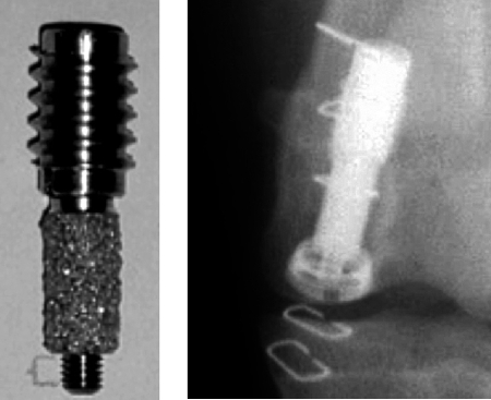
Weight-bearing implants with a 0.75-mm surrounding gap were inserted bilaterally in the knees of 10 dogs. Growth factors were incorporated in a biodegradable poly(D,L-lactide) coating on porous coated titanium implants. Plasma-sprayed HA implants served as controls. The dogs were killed at 4 weeks and the implants were evaluated by mechanical push-out test and by histomorphometry.
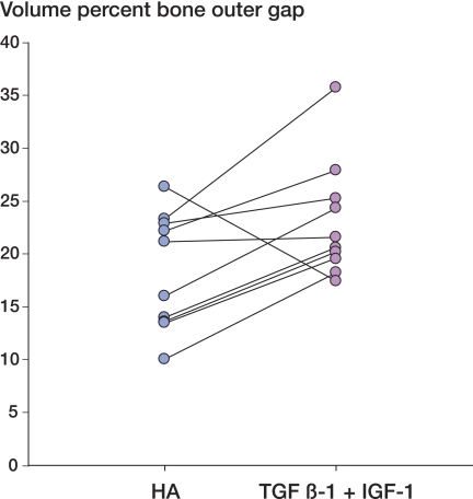
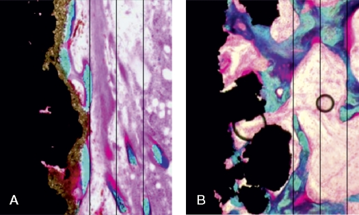
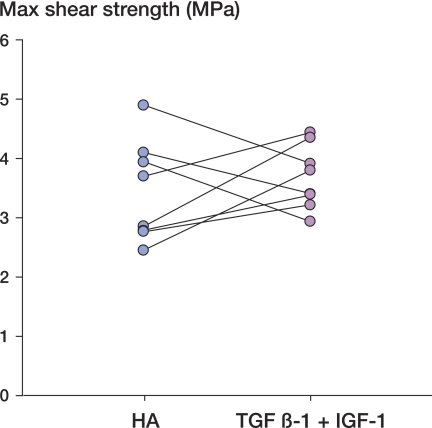
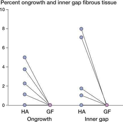
There was no difference in any of the mechanical parameters. Bone ongrowth was 3-fold higher for HA-coated implants (p < 0.001). For growth factor-coated implants, bone volume was 26% higher in the inner half of the gap and 28% higher in the outer half compared to HA (p < 0.03).
The mechanical fixation of porous-coated titanium implants with local growth factor release is comparable to that of HA coating. While HA mainly stimulated bone ongrowth, local release of TGF-beta1 and IGF-1 stimulated gap healing.
羟磷灰石(HA)涂层可刺激无水泥骨植入物的骨整合。最近,实验还表明局部释放成骨生长因子也可刺激骨整合,使骨骼填充骨科植入物周围的间隙。在此,我们比较了局部释放 TGF-β1 和 IGF-1 与 HA 涂层对植入物固定的影响。
将带有 0.75mm 周围间隙的负重植入物双侧插入 10 只狗的膝关节。将生长因子掺入多孔涂覆钛植入物的可生物降解聚(D,L-丙交酯)涂层中。等离子喷涂 HA 植入物作为对照。在 4 周时处死狗,并通过机械推出试验和组织形态计量学评估植入物。
在任何机械参数方面均无差异。HA 涂层植入物的骨长入增加了 3 倍(p<0.001)。对于生长因子涂层植入物,与 HA 相比,内半间隙的骨体积增加了 26%,外半间隙的骨体积增加了 28%(p<0.03)。
局部生长因子释放的多孔涂覆钛植入物的机械固定与 HA 涂层相当。虽然 HA 主要刺激骨长入,但局部释放 TGF-β1 和 IGF-1 刺激了间隙愈合。