Cvejic D, Selemetjev S, Savin S, Paunovic I, Tatic S
Institute for the Application of Nuclear Energy - INEP, University of Belgrade, Zemun - Belgrade, Serbia.
Eur J Histochem. 2009 Apr-Jun;53(2):e8. doi: 10.4081/ejh.2009.e8.
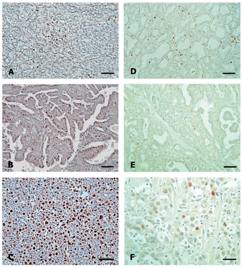
The aim of this study was to gain better insight into molecular changes which reflect disturbances in the balance between proliferation and apoptosis during progression of thyroid malignancy from papillary microcarcinoma (PMC) via clinically manifest papillary carcinoma (PTC) to anaplastic carcinoma (ATC). The apoptosis related molecules (Bcl-2, Bax) and proliferation related marker (PCNA) were analysed immunohistochemically in 120 archival cases comprising PMC (n=34), PTC (n=52) and ATC (n=34). In addition, in situ apoptotic cell death was analysed by the TUNEL method. The average Bcl-2 staining score did not differ between PMC and PTC (p>0.05), but was significantly lower in ATC (p<0.05).The Bax score was higher in PTCs and ATCs than in PMCs (p<0.05). Due to these changes, the Bcl-2/Bax ratio showed a marked decrease from PMC to ATC (p<0.05), while proliferation activity increased significantly from PTC to ATC (p<0.05). Despite high Bax expression, the rate of apoptotic cell death was low in the investigated carcinomas, especially in ATC, i.e. the increase in proliferative activity was not counterbalanced with appropriate cell death. Differences were found in the expression of apoptotic molecules (Bcl-2 and Bax), their ratio (Bcl-2 /Bax) and in the rate of apoptotic cell death and proliferative activity between PMC, PTC and ATC, indicating that disturbances in the balance between apoptosis and proliferation, in favour of the latter, occur gradually during the progression of malignancy in thyroid tumours.
本研究的目的是更深入地了解分子变化,这些变化反映了甲状腺恶性肿瘤从微小乳头状癌(PMC)经临床显性乳头状癌(PTC)发展为间变性癌(ATC)过程中增殖与凋亡平衡的紊乱。对120例存档病例进行免疫组织化学分析,检测凋亡相关分子(Bcl-2、Bax)和增殖相关标志物(PCNA),这些病例包括PMC(n = 34)、PTC(n = 52)和ATC(n = 34)。此外,采用TUNEL法分析原位凋亡细胞死亡情况。PMC和PTC之间的平均Bcl-2染色评分无差异(p>0.05),但ATC中的评分显著较低(p<0.05)。PTC和ATC中的Bax评分高于PMC(p<0.05)。由于这些变化,Bcl-2/Bax比值从PMC到ATC显著降低(p<0.05),而增殖活性从PTC到ATC显著增加(p<0.05)。尽管Bax表达较高,但在所研究的癌组织中,尤其是在ATC中,凋亡细胞死亡率较低,即增殖活性的增加未被适当的细胞死亡所抵消。在PMC、PTC和ATC之间,凋亡分子(Bcl-2和Bax)的表达、它们的比值(Bcl-2/Bax)、凋亡细胞死亡率和增殖活性存在差异,这表明在甲状腺肿瘤恶性进展过程中,凋亡与增殖平衡的紊乱逐渐发生,且更倾向于增殖。