Department of Cell and Developmental Biology, University of North Carolina, Chapel Hill, NC, USA.
Autophagy. 2009 Nov;5(8):1099-106. doi: 10.4161/auto.5.8.9825. Epub 2009 Nov 13.
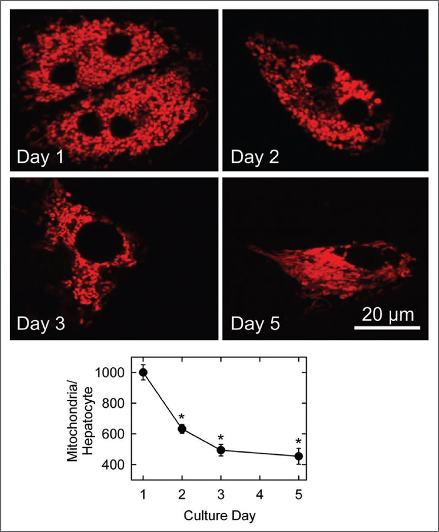
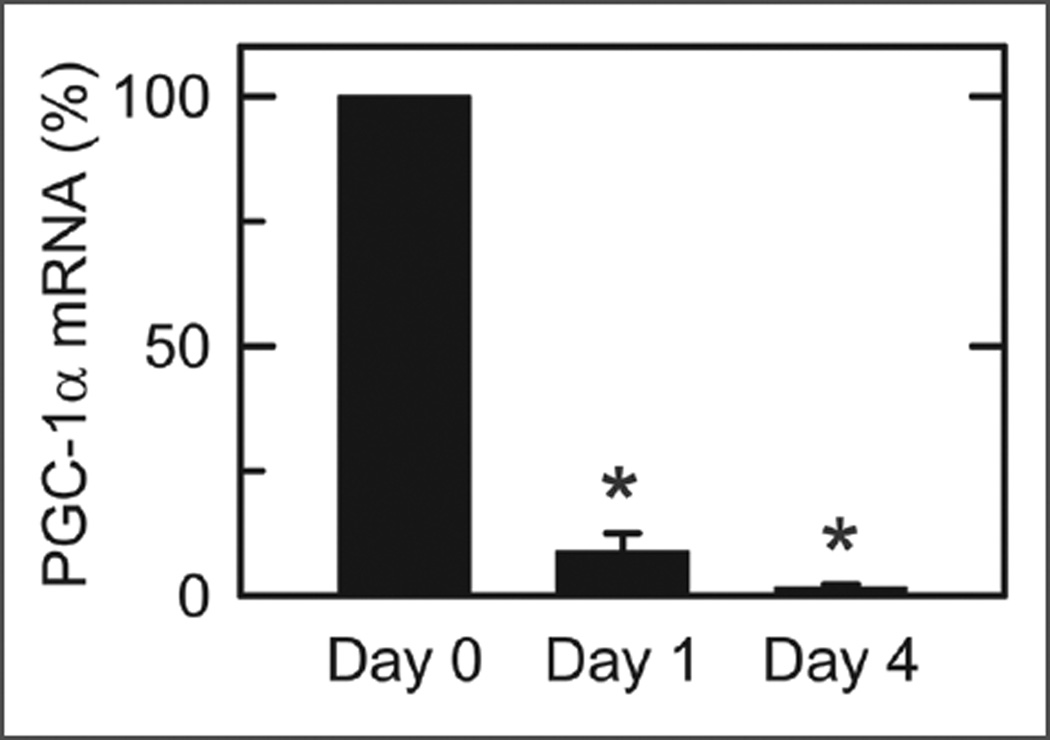
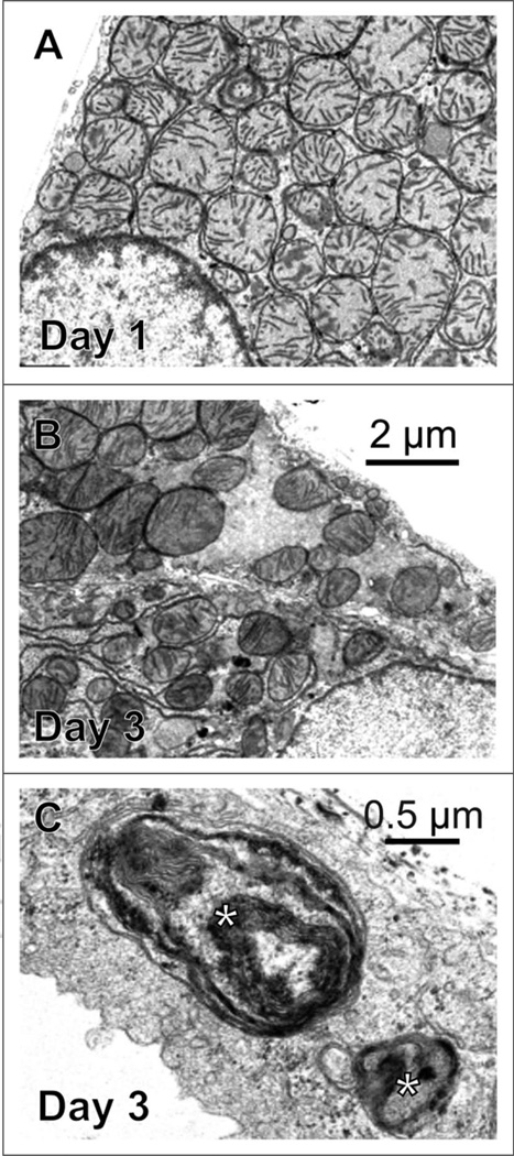
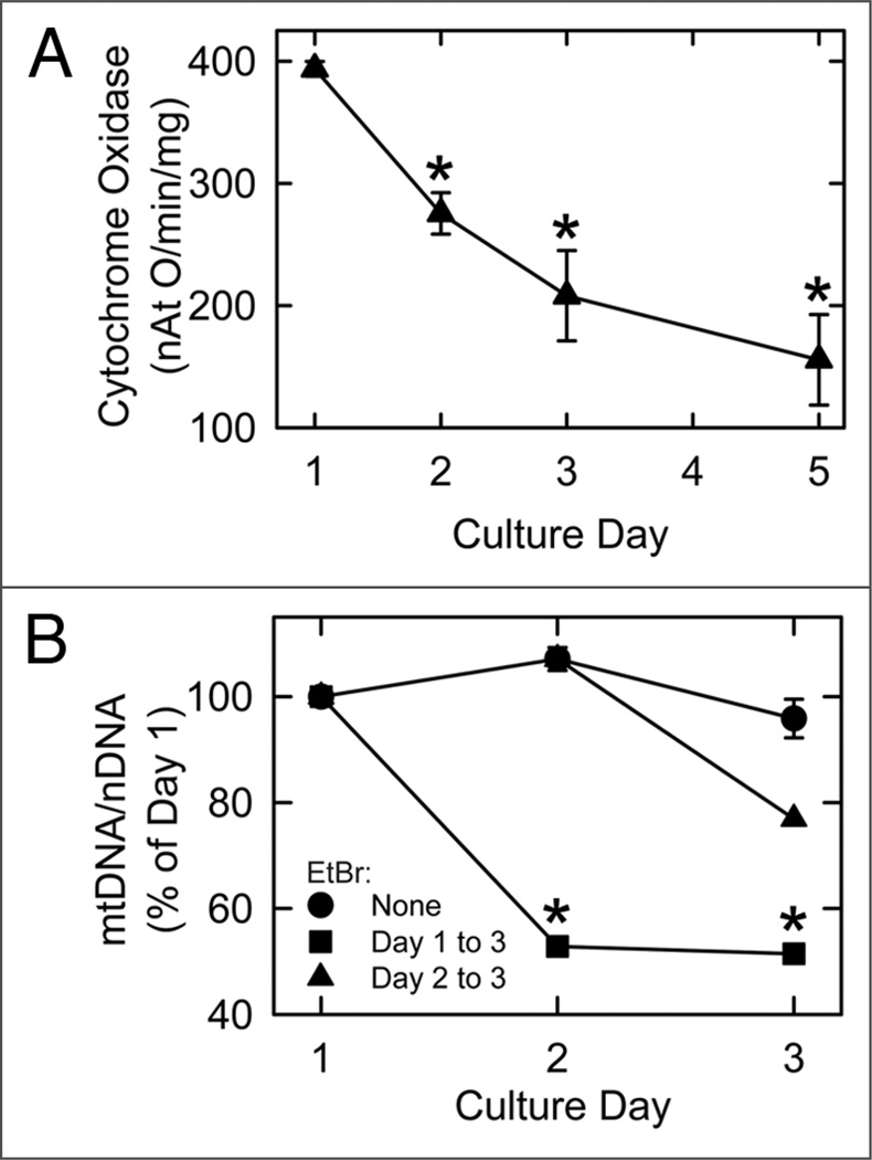
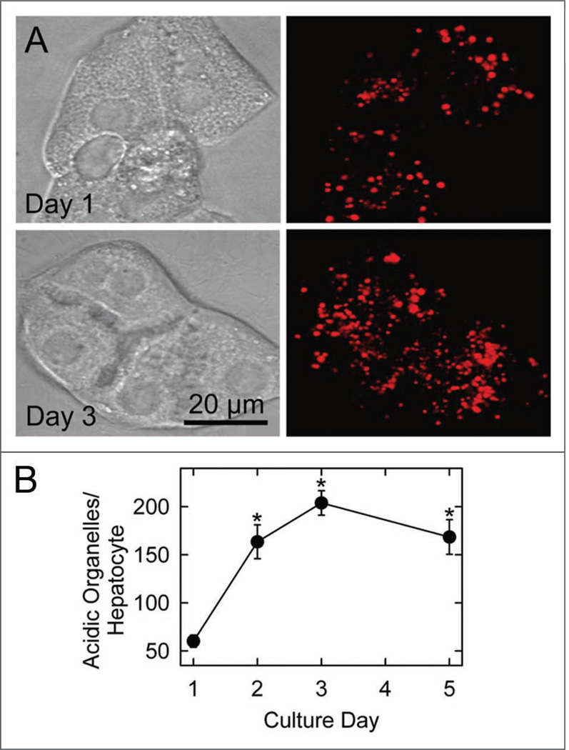
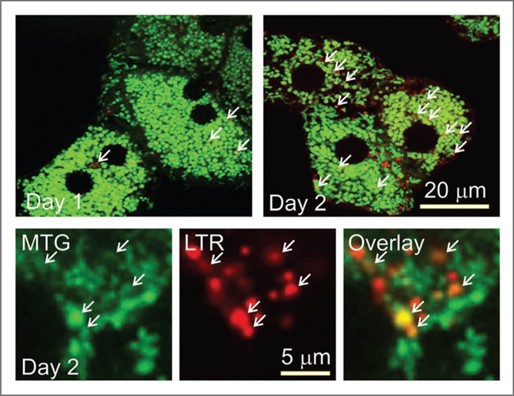
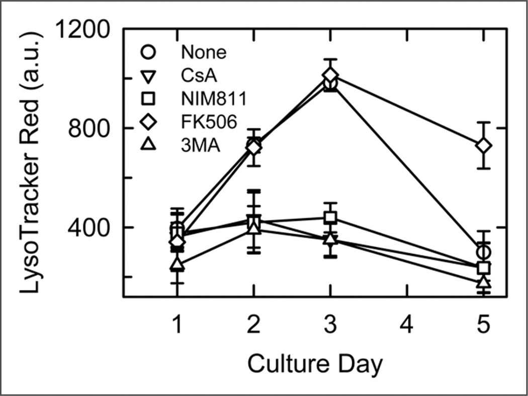
In primary culture, hepatocytes dedifferentiate, and their cytoplasm undergoes remodeling. Here, our aim was to characterize changes of mitochondria during remodeling. Hepatocytes were cultured one to five days in complete serumcontaining Waymouth's medium. In rat hepatocytes loaded with MitoTracker Green (MTG), tetramethylrhodamine methylester (TMRM), and/or LysoTracker Red (LTR), confocal microscopy revealed that mitochondria number and mass decreased by approximately 50% between Day 1 and Day 3 of culture. As mitochondria disappeared, lysosomes/autophagosomes proliferated five-fold. Decreased mitochondrial content correlated with (a) decreased cytochrome c oxidase activity and mitochondrial number observed by electron microscopy and (b) a profound decrease of PGC-1alpha mRNA expression. By contrast, mtDNA content per cell remained constant from the first to the third day of culture, although ethidium bromide (de novo mtDNA synthesis inhibitor) caused mtDNA to decrease by half from the first to the third culture day. As mitochondria disappeared, their MTG label moved into LTR-labeled lysosomes, which was indicative of autophagic degradation. A multiwell fluorescence assay revealed a 2.5-fold increase of autophagy on Day 3 of culture, which was decreased by 3-methyladenine, an inhibitor of autophagy, and also by cyclosporin A and NIM811, both selective inhibitors of the mitochondrial permeability transition (MPT). These findings indicate that mitochondrial autophagy (mitophagy) and the MPT underlie mitochondrial remodeling in cultured hepatocytes.
在原代培养中,肝细胞去分化,其细胞质发生重塑。在这里,我们的目的是研究重塑过程中线粒体的变化。将肝细胞在含有完全血清的 Waymouth 培养基中培养 1 至 5 天。在负载有 MitoTracker Green(MTG)、四甲基罗丹明甲酯(TMRM)和/或 LysoTracker Red(LTR)的大鼠肝细胞中,共聚焦显微镜显示,线粒体数量和质量在培养的第 1 天至第 3 天之间减少了约 50%。随着线粒体的消失,溶酶体/自噬体增殖了五倍。线粒体含量的减少与(a)电镜观察到的细胞色素 c 氧化酶活性和线粒体数量减少以及(b)PGC-1alpha mRNA 表达的显著减少相关。相比之下,尽管溴化乙锭(新合成 mtDNA 的抑制剂)使 mtDNA 在第 1 至第 3 天的培养中减少了一半,但从第 1 天到第 3 天,细胞内 mtDNA 含量保持不变。随着线粒体的消失,它们的 MTG 标记转移到 LTR 标记的溶酶体中,这表明发生了自噬降解。多孔荧光测定显示,培养第 3 天自噬增加了 2.5 倍,自噬被 3-甲基腺嘌呤(自噬抑制剂)、环孢菌素 A 和 NIM811(线粒体通透性转换(MPT)的选择性抑制剂)抑制。这些发现表明,线粒体自噬(mitophagy)和 MPT 是培养的肝细胞中线粒体重塑的基础。