Institute of Biomedical Engineering, National Yang-Ming University, Taiwan, ROC.
J Bone Miner Res. 2010 Mar;25(3):627-39. doi: 10.1359/jbmr.091008.
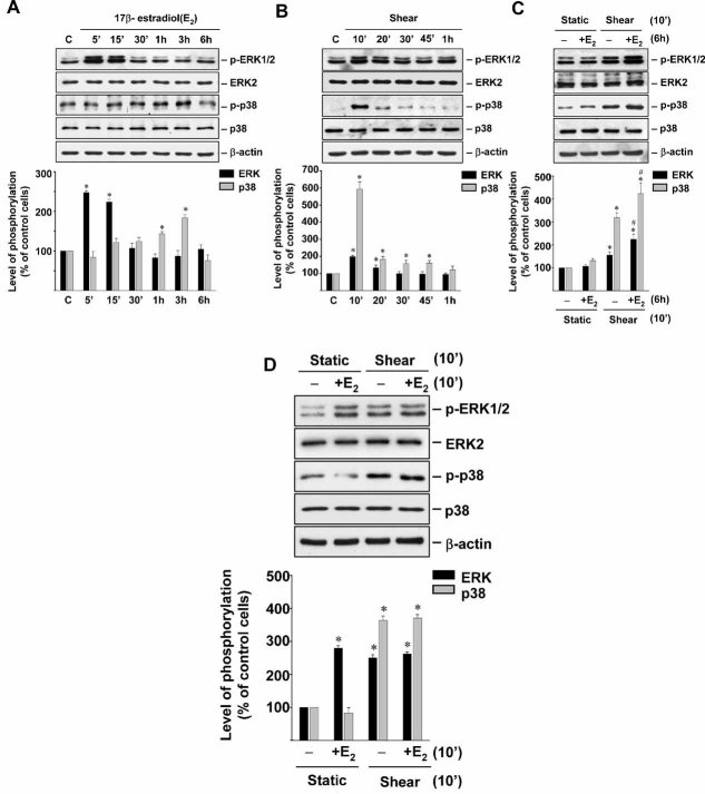
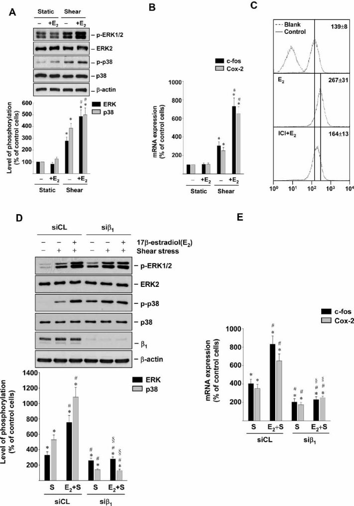
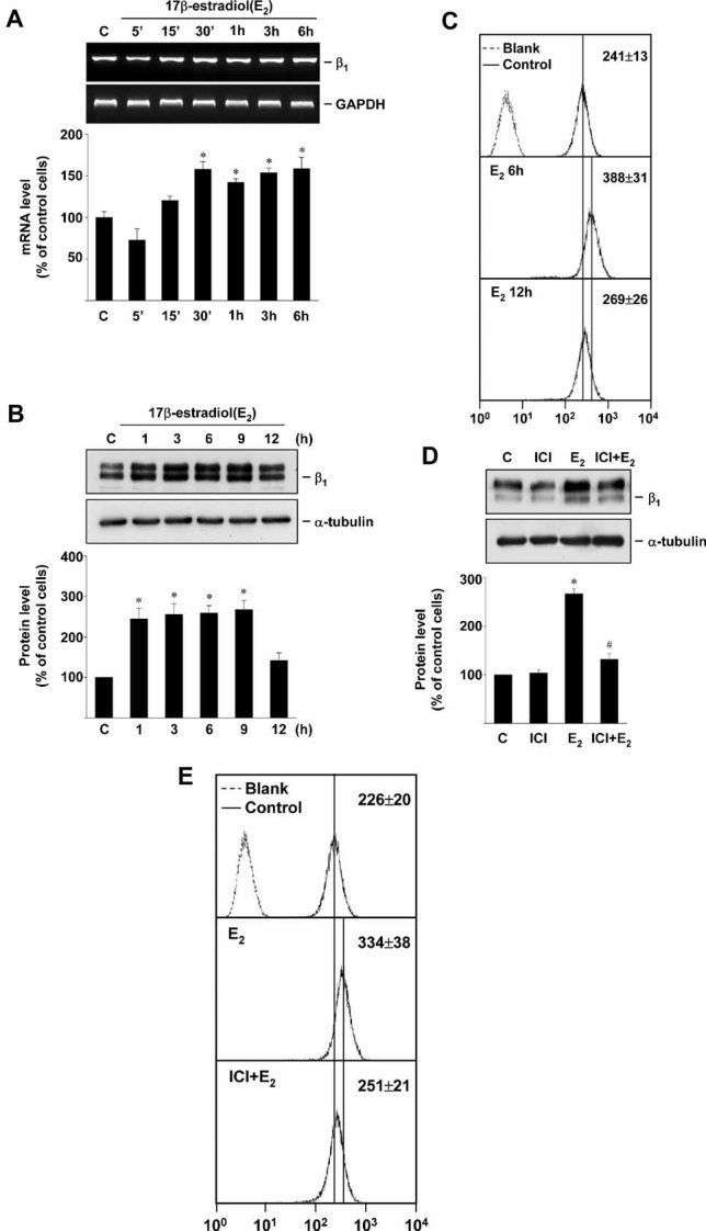
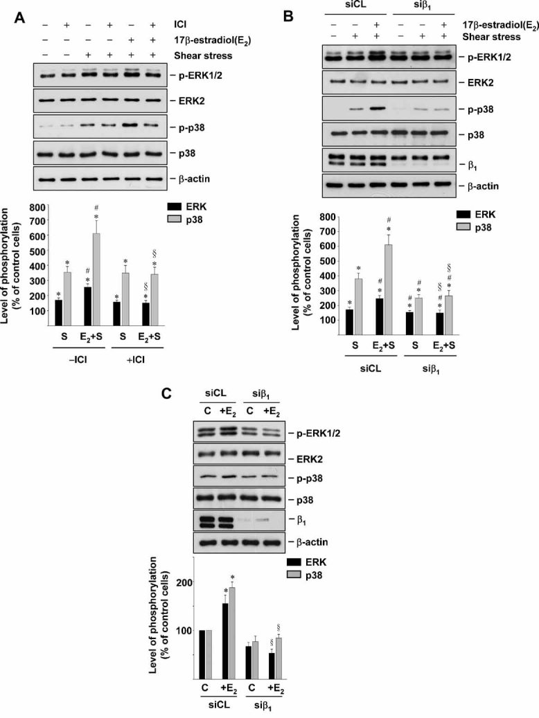
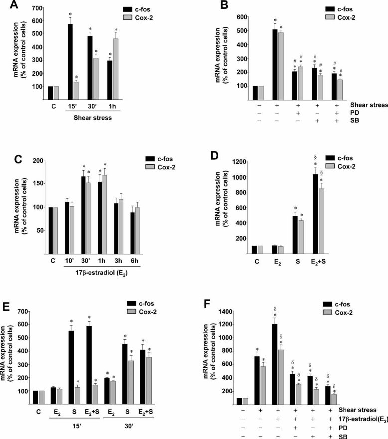
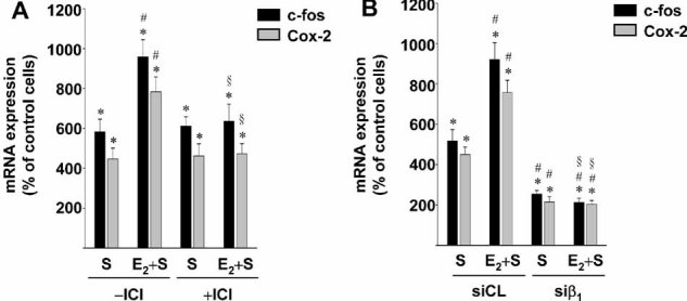
Estrogen and mechanical forces are positive regulators for osteoblast proliferation and bone formation. We investigated the synergistic effect of estrogen and flow-induced shear stress on signal transduction and gene expression in human osetoblast-like MG63 cells and primary osteoblasts (HOBs) using activations of extracellular signal-regulated kinase (ERK) and p38 mitogen-activated protein kinase (MAPK) and expressions of c-fos and cyclooxygenase-2 (I) as readouts. Estrogen (17beta-estradiol, 10 nM) and shear stress (12 dyn/cm(2)) alone induced transient phosphorylations of ERK and p38 MAPK in MG63 cells. Pretreating MG63 cells with 17beta-estradiol for 6 hours before shearing augmented these shear-induced MAPK phosphorylations. Western blot and flow cytometric analyses showed that treating MG63 cells with 17beta-estradiol for 6 hrs induced their beta(1)-integrin expression. This estrogen-induction of beta(1)-integrin was inhibited by pretreating the cells with a specific antagonist of estrogen receptor ICI 182,780. Both 17beta-estradiol and shear stress alone induced c-fos and Cox-2 gene expressions in MG63 cells. Pretreating MG63 cells with 17beta-estradiol for 6 hrs augmented the shear-induced c-fos and Cox-2 expressions. The augmented effects of 17beta-estradiol on shear-induced MAPK phosphorylations and c-fos and Cox-2 expressions were inhibited by pretreating the cells with ICI 182,780 or transfecting the cells with beta(1)-specific small interfering RNA. Similar results on the augmented effect of estrogen on shear-induced signaling and gene expression were obtained with HOBs. Our findings provide insights into the mechanism by which estrogen augments shear stress responsiveness of signal transduction and gene expression in bone cells via estrogen receptor-mediated increases in beta(1)-integrin expression.
雌激素和机械力是成骨细胞增殖和骨形成的正向调节剂。我们使用细胞外信号调节激酶 (ERK) 和丝裂原活化蛋白激酶 (p38 MAPK) 的激活以及 c-fos 和环氧化酶-2 (I) 的表达作为读出,研究了雌激素和流诱导切应力对人成骨样 MG63 细胞和原代成骨细胞 (HOBs) 的信号转导和基因表达的协同作用。雌激素 (17β-雌二醇,10 nM) 和切应力 (12 dyn/cm²) 单独诱导 MG63 细胞中 ERK 和 p38 MAPK 的瞬时磷酸化。在切变前用 17β-雌二醇预处理 MG63 细胞 6 小时增强了这些切变诱导的 MAPK 磷酸化。Western blot 和流式细胞术分析表明,用 17β-雌二醇处理 MG63 细胞 6 小时诱导其β1-整合素表达。用雌激素受体特异性拮抗剂 ICI 182,780 预处理细胞可抑制这种雌激素诱导的β1-整合素表达。17β-雌二醇和切应力单独诱导 MG63 细胞中 c-fos 和 Cox-2 基因表达。用 17β-雌二醇预处理 MG63 细胞 6 小时增强了切变诱导的 c-fos 和 Cox-2 表达。用 ICI 182,780 预处理细胞或转染β1-特异性小干扰 RNA 可抑制 17β-雌二醇对切变诱导的 MAPK 磷酸化和 c-fos 和 Cox-2 表达的增强作用。用 HOBs 获得了关于雌激素对切变诱导的信号转导和基因表达的增强作用的类似结果。我们的发现提供了关于雌激素通过雌激素受体介导的β1-整合素表达增加增强骨细胞中信号转导和基因表达对切变应激反应性的机制的见解。