Institute of Biomedicine, Department of Cell Biology and Anatomy, University of Turku, Finland.
BMC Cancer. 2009 Oct 12;9:362. doi: 10.1186/1471-2407-9-362.
Prostate cancer metastasizes to regional lymph nodes and distant sites but the roles of lymphatic and hematogenous pathways in metastasis are not fully understood.
We studied the roles of VEGF-C and VEGFR3 in prostate cancer metastasis by blocking VEGFR3 using intravenous adenovirus-delivered VEGFR3-Ig fusion protein (VEGFR3-Ig) and by ectopic expression of VEGF-C in PC-3 prostate tumors in nude mice.
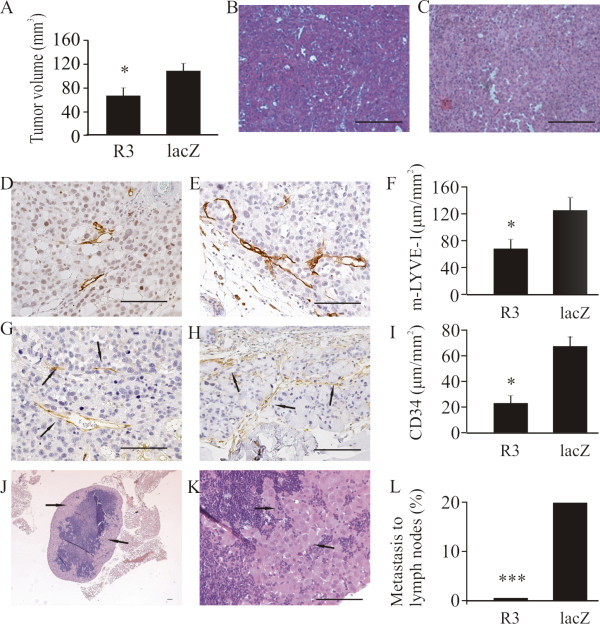
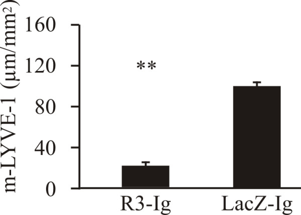
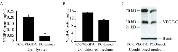
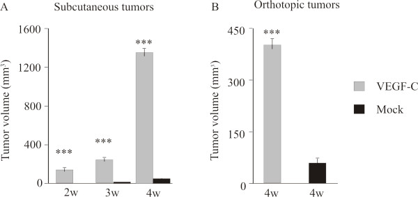
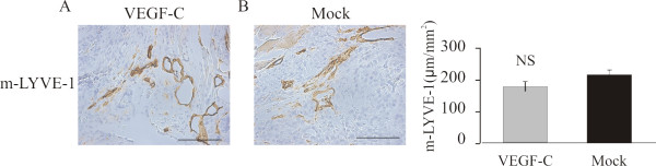
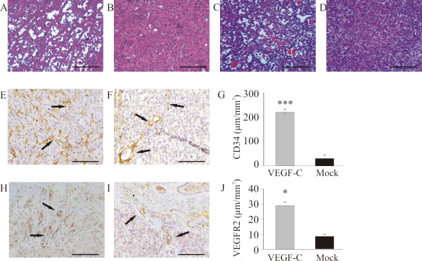
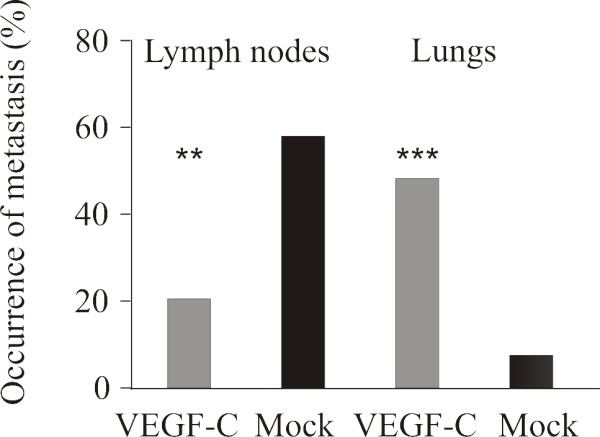
VEGFR3-Ig decreased the density of lymphatic capillaries in orthotopic PC-3 tumors (p < 0.05) and inhibited metastasis to iliac and sacral lymph nodes. In addition, tumor volumes were smaller in the VEGFR3-Ig-treated group compared with the control group (p < 0.05). Transfection of PC-3 cells with the VEGF-C gene led to a high level of 29/31 kD VEGF-C expression in PC-3 cells. The size of orthotopic and subcutaneous PC-3/VEGF-C tumors was significantly greater than that of PC-3/mock tumors (both p < 0.001). Interestingly, while most orthotopic PC-3 and PC-3/mock tumors grown for 4 weeks metastasized to prostate-draining lymph nodes, orthotopic PC-3/VEGF-C tumors primarily metastasized to the lungs. PC-3/VEGF-C tumors showed highly angiogenic morphology with an increased density of blood capillaries compared with PC-3/mock tumors (p < 0.001).
The data suggest that even though VEGF-C/VEGFR3 pathway is primarily required for lymphangiogenesis and lymphatic metastasis, an increased level of VEGF-C can also stimulate angiogenesis, which is associated with growth of orthotopic prostate tumors and a switch from a primary pattern of lymph node metastasis to an increased proportion of metastases at distant sites.
前列腺癌转移到局部淋巴结和远处部位,但淋巴和血行途径在转移中的作用尚不完全清楚。
我们通过静脉注射腺病毒介导的 VEGFR3-Ig 融合蛋白(VEGFR3-Ig)阻断 VEGFR3,以及在裸鼠 PC-3 前列腺肿瘤中异位表达 VEGF-C,研究了 VEGF-C 和 VEGFR3 在前列腺癌转移中的作用。
VEGFR3-Ig 降低了原位 PC-3 肿瘤中淋巴管的密度(p < 0.05),并抑制了髂骨和骶骨淋巴结的转移。此外,与对照组相比,VEGFR3-Ig 治疗组的肿瘤体积较小(p < 0.05)。PC-3 细胞中 VEGF-C 基因的转染导致 29/31 kD VEGF-C 在 PC-3 细胞中的高水平表达。原位和皮下 PC-3/VEGF-C 肿瘤的大小明显大于 PC-3/模拟肿瘤(均 p < 0.001)。有趣的是,虽然大多数 4 周生长的原位 PC-3 和 PC-3/模拟肿瘤转移到前列腺引流淋巴结,但原位 PC-3/VEGF-C 肿瘤主要转移到肺部。与 PC-3/模拟肿瘤相比,PC-3/VEGF-C 肿瘤表现出高度血管生成形态,血液毛细血管密度增加(p < 0.001)。
数据表明,尽管 VEGF-C/VEGFR3 途径主要用于淋巴管生成和淋巴转移,但 VEGF-C 水平的增加也可以刺激血管生成,这与原位前列腺肿瘤的生长以及从主要淋巴结转移模式向远处转移比例增加相关。