Wellman Center for Photomedicine, Massachusetts General Hospital, Harvard Medical School, Boston, MA 02114, USA.
Br J Cancer. 2009 Dec 15;101(12):2015-22. doi: 10.1038/sj.bjc.6605436. Epub 2009 Nov 17.
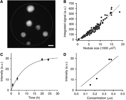
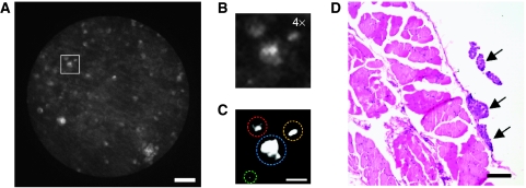
In patients with advanced ovarian cancer (OvCa), microscopic residual tumour nodules that remain after surgical debulking frequently escape detection by current treatment assessment methods and lead to disease recurrence. The aim of this study was to evaluate the use of high-resolution fibre-optic fluorescence imaging of the clinically approved photodynamic therapy (PDT) agent benzoporphyin-derivative monoacid ring A (BPD-MA) for detection of microscopic OvCa and for monitoring treatment response.
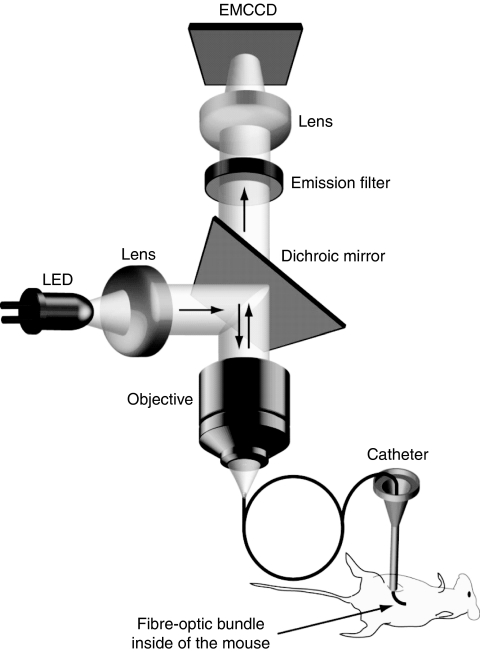
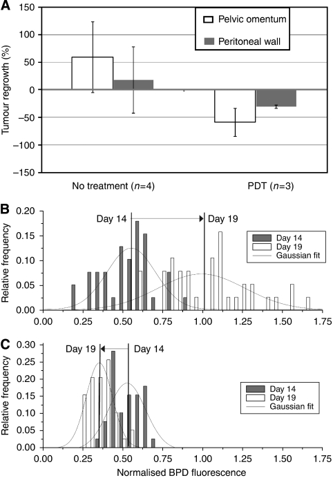
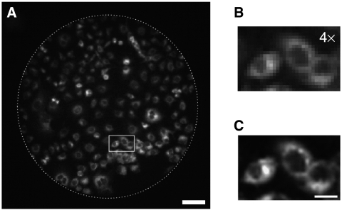
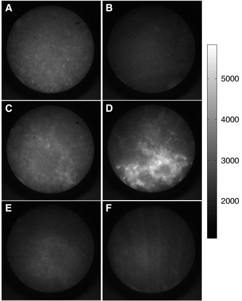
Our fluorescence microendoscope consists of a flexible imaging fibre coupled to a custom epi-fluorescence system optimised for imaging BPD-MA, which, after a single administration, serves as both an imaging agent and a light-activated therapeutic agent. After characterisation in an in vitro OvCa 3D model, we used the flexible imaging fibre to minimally invasively image the peritoneal cavity of a disseminated OvCa murine model using BPD-MA administered intraperitoneally (i.p.). To evaluate longitudinal changes in response to treatment, we compared sets of images obtained before and after PDT with those from untreated mice imaged at the same time points.
By comparison with histopathology, we report an 86% sensitivity for tumour detection in vivo using the microendoscope. Using a custom routine to batch process-image data in the monitoring study, treated mice exhibited an average decrease of 58.8% in tumour volumes compared with an increase of 59.3% in untreated controls (P<0.05).
Our findings indicate the potential of this approach as a reporter of treatment outcome that could aid in the rational design of strategies to mitigate recurrent OvCa.
在晚期卵巢癌(OvCa)患者中,手术减瘤后残留的微小肿瘤结节经常逃脱当前治疗评估方法的检测,导致疾病复发。本研究旨在评估临床批准的光动力疗法(PDT)药物苯并卟啉衍生物单酸环 A(BPD-MA)的高分辨率光纤荧光成像在检测微小 OvCa 和监测治疗反应中的应用。
我们的荧光显微镜由一根柔性成像光纤与一个定制的 epi-fluorescence 系统耦合而成,该系统经过优化可用于成像 BPD-MA,在单次给药后,它既是成像剂又是光激活治疗剂。在体外 OvCa 3D 模型中进行特征描述后,我们使用柔性成像光纤通过腹腔内(i.p.)给予 BPD-MA 对弥散性 OvCa 小鼠模型的腹腔进行微创成像。为了评估对治疗的纵向变化,我们将 PDT 前后获得的图像与同一时间点未治疗小鼠的图像进行了比较。
与组织病理学相比,我们报告了体内使用显微镜检测肿瘤的灵敏度为 86%。在监测研究中,使用自定义例程批量处理图像数据,与未治疗的对照组相比,治疗组的肿瘤体积平均减少了 58.8%,而对照组的肿瘤体积增加了 59.3%(P<0.05)。
我们的研究结果表明,这种方法具有作为治疗结果报告的潜力,可能有助于合理设计减轻复发性 OvCa 的策略。