Department of Genitourinary Medical Oncology Research, The University of Texas M.D. Anderson Cancer Center, Houston, TX, USA.
Cancer Biol Ther. 2009 Dec;8(23):2286-96. doi: 10.4161/cbt.8.23.10138. Epub 2009 Dec 19.
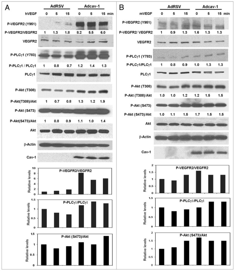
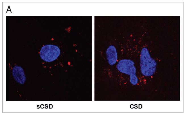
Caveolin-1 (cav-1) is a multifunctional protein and major component of caveolae membranes serving important functions related to signal transduction, endocytosis, transcytosis, and molecular transport. We previously showed that cav-1 is overexpressed and secreted by metastatic prostate cancer cells. We now report that cav-1 gene transduction (Adcav-1) or recombinant cav-1 (rcav-1) protein treatment of cav-1-negative prostate cancer cell line LP-LNCaP or cav-1(-/-) endothelial cells potentiated VEGF-stimulated angiogenic signaling. Downregulation of cav-1 in prostate cancer cell line PC-3 or human umbilical vein endothelial cells (HUVECs) through cav-1 siRNA significantly reduced basal and VEGF-stimulated phosphorylation of VEGFR2 (Y951), PLCgamma1 (Y783) and/or Akt (S473 & T308) relative to those in control siRNA treated cells. Additionally rcav-1 stimulation of cav-1 siRNA treated HUVECs restored this signaling pathway. Confocal microscopy and immunoprecipitation analysis revealed association and colocalization of VEGFR2 and PLCgamma1 with cav-1 following VEGF stimulation in HUVECs. Interestingly, treatment of HUVECs with cav-1 scaffolding domain (CSD) caused significant reduction in the VEGF-stimulated phosphorylation of VEGFR2, PLCgamma1 and Akt suggesting that CSD inhibits cav-1-mediated angiogenic signaling. VEGF stimulation of HUVECs significantly increased tubule length and cell migration, but this stimulatory effect was significantly reduced by cav-1 siRNA and/or CSD treatment. The present study demonstrates that cav-1 regulates VEGF-stimulated VEGFR2 autophosphorylation and activation of downstream angiogenic signaling, possibly through compartmentalization of specific signaling molecules. Our results provide mechanistic insight into the role of cav-1 in prostate cancer and suggest the use of CSD as a therapeutic tool to suppress angiogenic signaling in prostate cancer.
窖蛋白-1(cav-1)是一种多功能蛋白,是质膜窖的主要组成部分,具有重要的信号转导、内吞作用、转胞吞作用和分子运输功能。我们之前曾报道过转移性前列腺癌细胞过表达和分泌窖蛋白-1。现在我们报告说,窖蛋白-1基因转导(Adcav-1)或重组窖蛋白-1(rcav-1)蛋白处理窖蛋白-1阴性前列腺癌细胞系 LP-LNCaP 或窖蛋白-1(-/-)内皮细胞增强了 VEGF 刺激的血管生成信号。通过窖蛋白-1 siRNA下调前列腺癌细胞系 PC-3 或人脐静脉内皮细胞(HUVEC)中的窖蛋白-1,与对照 siRNA 处理的细胞相比,显著降低了基础状态和 VEGF 刺激的 VEGFR2(Y951)、PLCγ1(Y783)和/或 Akt(S473 和 T308)的磷酸化。此外,rcav-1 刺激窖蛋白-1 siRNA 处理的 HUVEC 恢复了这条信号通路。共聚焦显微镜和免疫沉淀分析显示,在 HUVEC 中 VEGF 刺激后,VEGFR2 和 PLCγ1 与窖蛋白-1 结合并共定位。有趣的是,用窖蛋白-1支架结构域(CSD)处理 HUVEC 会导致 VEGF 刺激的 VEGFR2、PLCγ1 和 Akt 的磷酸化显著减少,这表明 CSD 抑制了窖蛋白-1 介导的血管生成信号。VEGF 刺激 HUVEC 显著增加了管腔长度和细胞迁移,但这种刺激作用被窖蛋白-1 siRNA 和/或 CSD 处理显著降低。本研究表明,窖蛋白-1 调节 VEGF 刺激的 VEGFR2 自身磷酸化和下游血管生成信号的激活,可能是通过特定信号分子的区室化。我们的结果提供了对窖蛋白-1 在前列腺癌中的作用的机制见解,并表明使用 CSD 作为一种治疗工具来抑制前列腺癌中的血管生成信号。