CNRS, UMR 8620, Orsay, France.
PLoS One. 2009 Nov 19;4(11):e7901. doi: 10.1371/journal.pone.0007901.
Physical exercise has been shown to increase adult neurogenesis in the dentate gyrus and enhances synaptic plasticity. The antiapoptotic kinase, Akt has also been shown to be phosphorylated following voluntary exercise; however, it remains unknown whether the PI3K-Akt signaling pathway is involved in exercise-induced neurogenesis and the associated facilitation of synaptic plasticity in the dentate gyrus.
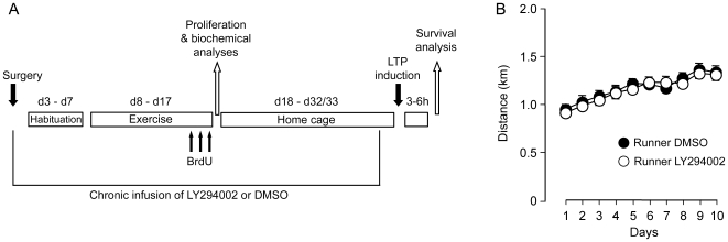
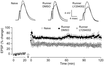
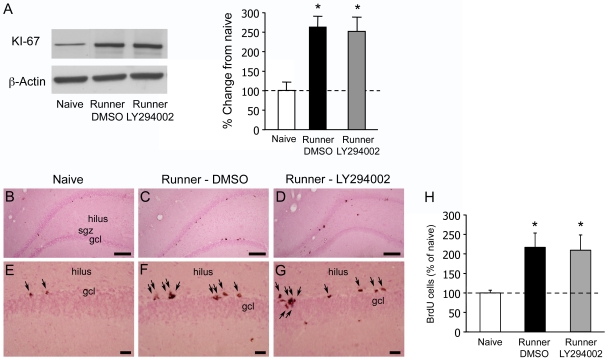
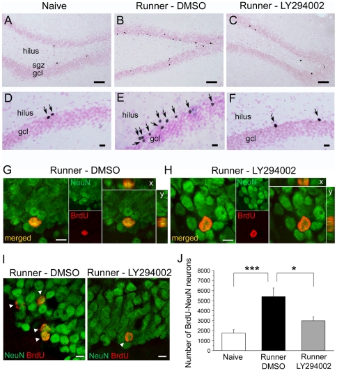
METHODOLOGY/PRINCIPAL FINDINGS: To gain insight into the potential role of this signaling pathway in exercise-induced neurogenesis and LTP in the dentate gyrus rats were infused with the PI3K inhibitor, LY294002 or vehicle control solution (icv) via osmotic minipumps and exercised in a running wheel for 10 days. Newborn cells in the dentate gyrus were date-labelled with BrdU on the last 3 days of exercise. Then, they were either returned to the home cage for 2 weeks to assess exercise-induced LTP and neurogenesis in the dentate gyrus, or were killed on the last day of exercise to assess proliferation and activation of the PI3K-Akt cascade using western blotting.
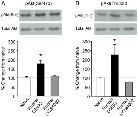
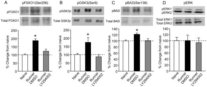
CONCLUSIONS/SIGNIFICANCE: Exercise increases cell proliferation and promotes survival of adult-born neurons in the dentate gyrus. Immediately after exercise, we found that Akt and three downstream targets, BAD, GSK3beta and FOXO1 were activated. LY294002 blocked exercise-induced phosphorylation of Akt and downstream target proteins. This had no effect on exercise-induced cell proliferation, but it abolished most of the beneficial effect of exercise on the survival of newly generated dentate gyrus neurons and prevented exercise-induced increase in dentate gyrus LTP. These results suggest that activation of the PI3 kinase-Akt signaling pathway plays a significant role via an antiapoptotic function in promoting survival of newly formed granule cells generated during exercise and the associated increase in synaptic plasticity in the dentate gyrus.
运动已被证明可以增加齿状回中的成年神经发生,并增强突触可塑性。抗细胞凋亡激酶 Akt 在进行自愿运动后也被证明会被磷酸化;然而,目前尚不清楚 PI3K-Akt 信号通路是否参与运动诱导的神经发生以及与齿状回中突触可塑性的相关促进作用。
方法/主要发现:为了深入了解该信号通路在运动诱导的神经发生和齿状回中的长时程增强(LTP)中的潜在作用,通过渗透微型泵向大鼠脑室内输注 PI3K 抑制剂 LY294002 或载体对照溶液(icv),并在跑步轮中运动 10 天。在运动的最后 3 天,用 BrdU 对齿状回中的新生细胞进行标记。然后,它们要么返回普通笼中 2 周以评估齿状回中的运动诱导的 LTP 和神经发生,要么在运动的最后一天处死以评估 PI3K-Akt 级联的增殖和激活情况,使用 Western blot 进行检测。
结论/意义:运动可增加细胞增殖并促进齿状回中成年新生神经元的存活。运动后立即,我们发现 Akt 和三个下游靶标 BAD、GSK3β 和 FOXO1 被激活。LY294002 阻断了 Akt 和下游靶蛋白的运动诱导磷酸化。这对运动诱导的细胞增殖没有影响,但它消除了运动对新生成的齿状回神经元存活的大部分有益影响,并防止了运动诱导的齿状回 LTP 增加。这些结果表明,PI3 激酶-Akt 信号通路的激活通过抗凋亡功能在促进运动过程中产生的新颗粒细胞的存活以及与齿状回中突触可塑性的相关增加方面发挥着重要作用。