Lehrstuhl für Zellbiologie, Universität Konstanz, Konstanz, Germany.
BMC Biol. 2009 Nov 25;7:81. doi: 10.1186/1741-7007-7-81.
Bacteria-triggered signaling events in infected host cells are key elements in shaping the host response to pathogens. Within the eukaryotic cell, signaling complexes are spatially organized. However, the investigation of protein-protein interactions triggered by bacterial infection in the cellular context is technically challenging. Here, we provide a methodological approach to exploit fluorescence resonance energy transfer (FRET) to visualize pathogen-initiated signaling events in human cells.
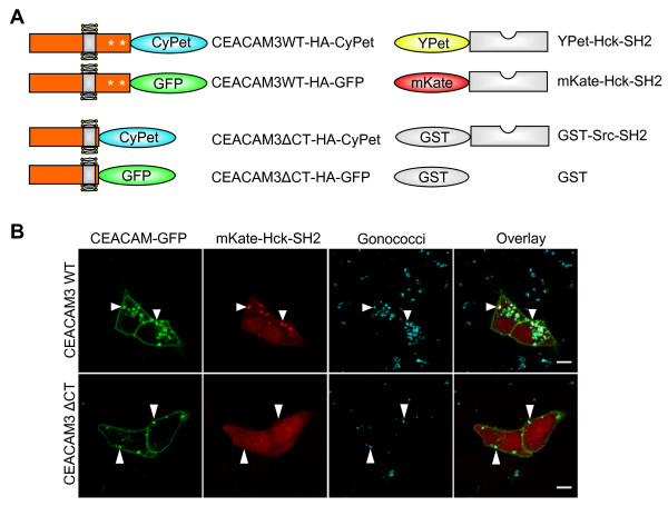
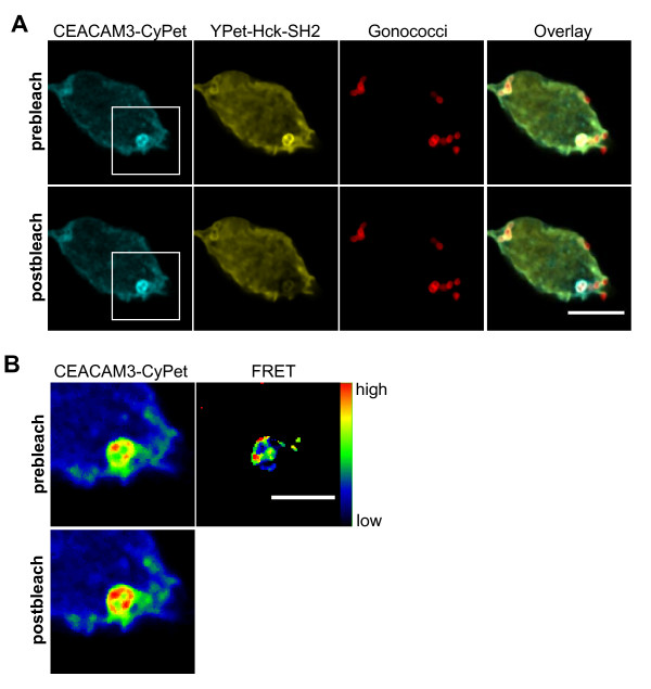
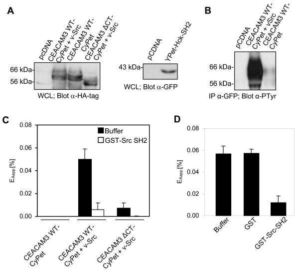
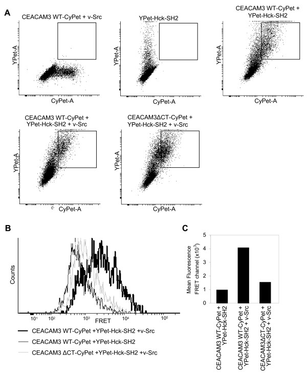
Live-cell microscopy revealed the transient recruitment of the Src family tyrosine kinase Hck upon bacterial engagement of the receptor carcinoembryonic antigen-related cell adhesion molecule 3 (CEACAM3). In cells expressing a CEACAM3 variant lacking the cytoplasmic domain, the Src homology 2 (SH2) domain of Hck (Hck-SH2) was not recruited, even though bacteria still bound to the receptor. FRET measurements on the basis of whole cell lysates revealed intimate binding between Hck-SH2 (using enhanced yellow fluorescent protein (YPet)-Hck-SH2) and the tyrosine-phosphorylated enhanced cyan fluorescent protein-labeled cytoplasmic domain of wild-type CEACAM3 (CEACAM3 WT-CyPet) and a flow cytometry-based FRET approach verified this association in intact cells. Using confocal microscopy and acceptor photobleaching, FRET between Hck-SH2 and CEACAM3 was localized to the sites of bacteria-host cell contact.
These data demonstrate not only the intimate binding of the SH2 domain of Hck to the tyrosine-phosphorylated cytoplasmic domain of CEACAM3 in intact cells, but furthermore, FRET measurements allow the subcellular localization of this process during bacterial infection. FRET-based assays are valuable tools to resolve bacteria-induced protein-protein interactions in the context of the intact host cell.
感染宿主细胞中细菌触发的信号事件是宿主对病原体反应的关键因素。在真核细胞内,信号复合物是空间组织的。然而,在细胞环境中研究细菌感染引发的蛋白质-蛋白质相互作用在技术上具有挑战性。在这里,我们提供了一种方法学方法,利用荧光共振能量转移(FRET)来可视化人类细胞中病原体引发的信号事件。
活细胞显微镜显示,在细菌与受体癌胚抗原相关细胞粘附分子 3(CEACAM3)结合后,Src 家族酪氨酸激酶 Hck 会短暂募集。在表达缺乏细胞质结构域的 CEACAM3 变体的细胞中,即使细菌仍与受体结合,Hck 的 Src 同源 2(SH2)结构域(Hck-SH2)也不会募集。基于全细胞裂解物的 FRET 测量显示 Hck-SH2(使用增强型黄色荧光蛋白(YPet)-Hck-SH2)与野生型 CEACAM3 的酪氨酸磷酸化增强型青色荧光蛋白标记的细胞质结构域(CEACAM3 WT-CyPet)之间存在紧密结合,并且基于流式细胞术的 FRET 方法在完整细胞中验证了这种关联。使用共聚焦显微镜和受体光漂白,Hck-SH2 和 CEACAM3 之间的 FRET 定位于细菌-宿主细胞接触部位。
这些数据不仅证明了 Hck 的 SH2 结构域与完整细胞中 CEACAM3 的酪氨酸磷酸化细胞质结构域的紧密结合,而且 FRET 测量允许在细菌感染过程中对该过程进行亚细胞定位。FRET 基测定是解决完整宿主细胞中细菌诱导的蛋白质-蛋白质相互作用的有价值的工具。