Department of Podiatry, Faculty of Health Sciences, La Trobe University, Bundoora, Australia.
J Foot Ankle Res. 2009 Nov 26;2:35. doi: 10.1186/1757-1146-2-35.
Some studies have found that flat-arched foot posture is related to altered lower limb muscle function compared to normal- or high-arched feet. However, the results from these studies were based on highly selected populations such as those with rheumatoid arthritis. Therefore, the objective of this study was to compare lower limb muscle function of normal and flat-arched feet in people without pain or disease.
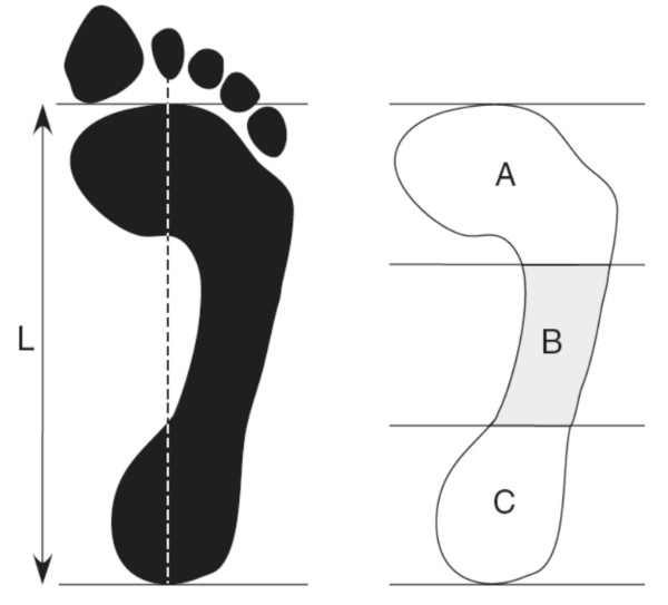
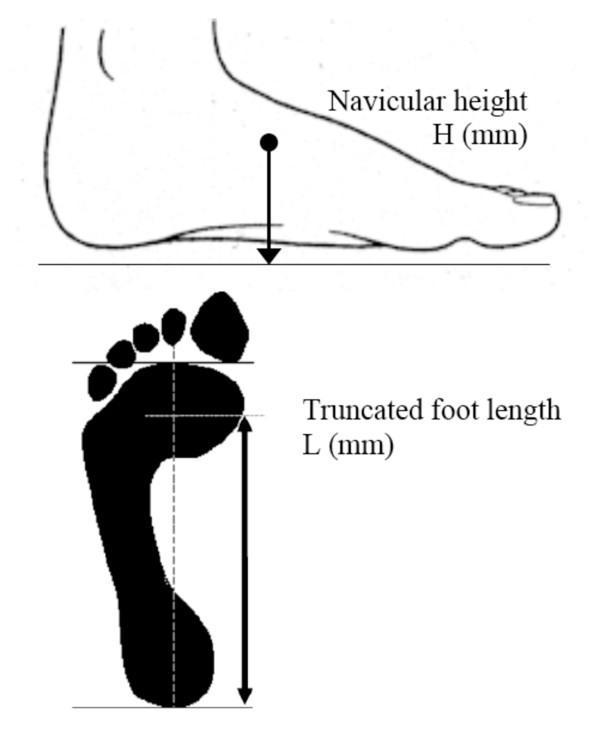
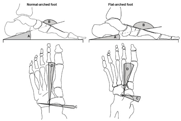
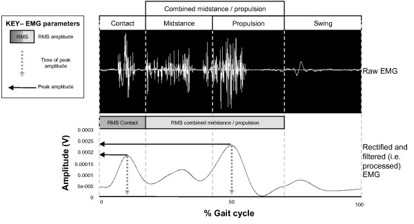
Sixty adults aged 18 to 47 years were recruited to this study. Of these, 30 had normal-arched feet (15 male and 15 female) and 30 had flat-arched feet (15 male and 15 female). Foot posture was classified using two clinical measurements (the arch index and navicular height) and four skeletal alignment measurements from weightbearing foot x-rays. Intramuscular fine-wire electrodes were inserted into tibialis posterior and peroneus longus under ultrasound guidance, and surface EMG activity was recorded from tibialis anterior and medial gastrocnemius while participants walked barefoot at their self-selected comfortable walking speed. Time of peak amplitude, peak and root mean square (RMS) amplitude were assessed from stance phase EMG data. Independent samples t-tests were performed to assess for significant differences between the normal- and flat-arched foot posture groups.
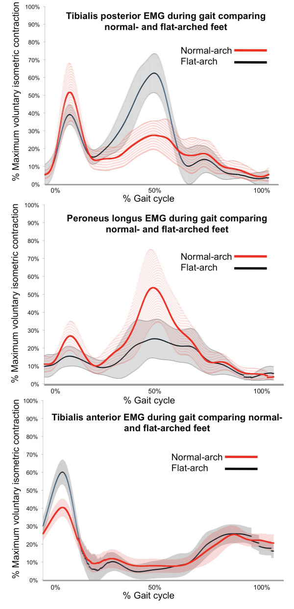
During contact phase, the flat-arched group exhibited increased activity of tibialis anterior (peak amplitude; 65 versus 46% of maximum voluntary isometric contraction) and decreased activity of peroneus longus (peak amplitude; 24 versus 37% of maximum voluntary isometric contraction). During midstance/propulsion, the flat-arched group exhibited increased activity of tibialis posterior (peak amplitude; 86 versus 60% of maximum voluntary isometric contraction) and decreased activity of peroneus longus (RMS amplitude; 25 versus 39% of maximum voluntary isometric contraction). Effect sizes for these significant findings ranged from 0.48 to 1.3, representing moderate to large differences in muscle activity between normal-arched and flat-arched feet.
Differences in muscle activity in people with flat-arched feet may reflect neuromuscular compensation to reduce overload of the medial longitudinal arch. Further research is required to determine whether these differences in muscle function are associated with injury.
一些研究发现,与正常足或高足弓相比,扁平足足弓的足部姿势与下肢肌肉功能的改变有关。然而,这些研究的结果是基于高度选择的人群,如类风湿关节炎患者。因此,本研究的目的是比较无疼痛或疾病人群中正常足弓和扁平足弓的下肢肌肉功能。
本研究共招募了 60 名 18 至 47 岁的成年人。其中,30 名受试者有正常足弓(15 名男性和 15 名女性),30 名受试者有扁平足弓(15 名男性和 15 名女性)。足部姿势通过两种临床测量(足弓指数和舟骨高度)和负重足部 X 线的四个骨骼对线测量来分类。在超声引导下将肌内细导线电极插入比目鱼肌和腓骨长肌,并在受试者以自我选择的舒适步行速度赤脚行走时记录胫骨前肌和内侧腓肠肌的表面肌电图活动。从站立相肌电图数据评估峰值幅度、峰值和均方根(RMS)幅度的时间。使用独立样本 t 检验评估正常足弓和扁平足弓姿势组之间的显著差异。
在接触阶段,扁平足组表现出胫骨前肌活动增加(峰值幅度;65%与最大等长收缩的 46%)和腓骨长肌活动减少(峰值幅度;24%与最大等长收缩的 37%)。在中足/推进阶段,扁平足组表现出胫骨后肌活动增加(峰值幅度;86%与最大等长收缩的 60%)和腓骨长肌活动减少(RMS 幅度;25%与最大等长收缩的 39%)。这些显著发现的效应大小范围为 0.48 至 1.3,代表正常足弓和扁平足弓之间肌肉活动的差异较大。
扁平足患者肌肉活动的差异可能反映了神经肌肉的代偿,以减少内侧纵弓的过载。需要进一步研究这些肌肉功能的差异是否与损伤有关。