Dipartimento di Genetica e Biologia Molecolare, Sapienza Università di Roma, Italy.
J Exp Clin Cancer Res. 2009 Dec 11;28(1):151. doi: 10.1186/1756-9966-28-151.
Many experimental data evidence that over-expression of various growth factors cause disorders in cell proliferation. The role of the Fibroblast Growth Factors (FGF) in growth control is indisputable: in particular, FGF1 and its tyrosine kinase receptor (FGFR1) act through a very complex network of mechanisms and pathways. In this work we have evaluated the antiproliferative activity effect of PD166866, a synthetic molecule inhibiting the tyrosin kinase action of FGFR1.
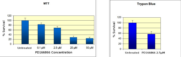
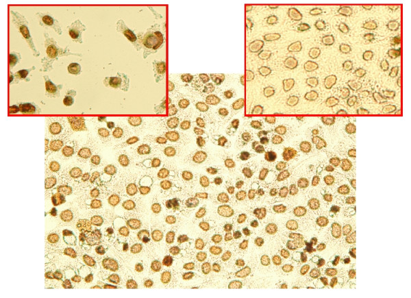
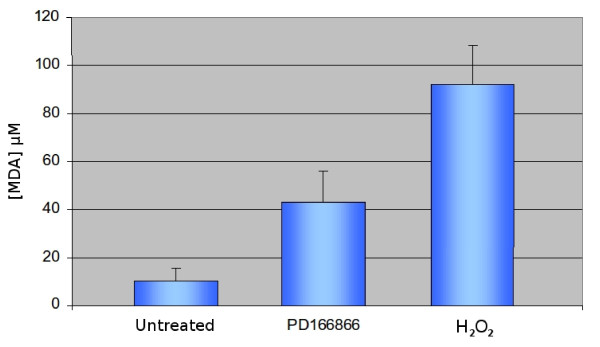
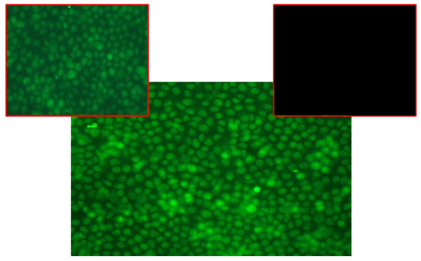
Cells were routinely grown in Dulbecco Modified Eagle's medium supplemented with newborn serum and a penicillin-streptomycin mixture.Cell viability was evaluated by Mosmann assay and by trypan blue staining. DNA damage was assessed by in situ fluorescent staining with Terminal Deoxynucleotidyl Transferase dUTP nick end labeling (TUNEL assay).Assessment of oxidative stress at membrane level was measured by quantitative analysis of the intra-cellular formation of malonyl-dialdheyde (MDA) deriving from the decomposition of poly-unsaturated fatty acids.The expression of Poly-ADP-Ribose-Polymerase (PARP), consequent to DNA fragmentation, was evidenced by immuno-histochemistry utilizing an antibody directed against an N-terminal fragment of the enzyme.
The bioactivity of the drug was investigated on Hela cells. Cytoxicity was assessed by the Mosmann assay and by vital staining with trypan blue. The target of the molecule is most likely the cell membrane as shown by the significant increase of the intracellular concentration of malonyl-dihaldheyde. The increase of this compound, as a consequence of the treatment with PD166866, is suggestive of membrane lipoperoxidation. The TUNEL assay gave a qualitative, though clear, indication of DNA damage. Furthermore we demonstrate intracellular accumulation of poly-ADP-ribose polymerase I. This enzyme is a sensor of nicks on the DNA strands and this supports the idea that treatment with the drug induces cell death.
Data presented in this work show that PD166866 has clear antiproliferative effects. The negative control of cell proliferation may be exerted through the activation of the apoptotic pathway. The results of experiments addressing this specific point, such as: evaluation of DNA damage, lipoperoxidation of the cell membrane and increase of expression of PARP, an enzyme directly involved in DNA repair. Results suggest that cells exposed to PD16866 undergo apoptosis. However, concomitant modes of cell death cannot be ruled out. The possible use of this drug for therapeutic purposes is discussed.
许多实验数据表明,各种生长因子的过度表达会导致细胞增殖紊乱。成纤维细胞生长因子(FGF)在生长控制中的作用是不可争议的:特别是,FGF1 及其酪氨酸激酶受体(FGFR1)通过一个非常复杂的机制和途径网络发挥作用。在这项工作中,我们评估了 PD166866 的抗增殖活性,PD166866 是一种抑制 FGFR1 酪氨酸激酶活性的合成分子。
细胞常规在添加新生血清和青霉素-链霉素混合物的 Dulbecco 改良 Eagle 培养基中生长。通过 Mosmann 测定法和台盼蓝染色评估细胞活力。通过原位荧光染色用末端脱氧核苷酸转移酶 dUTP 缺口末端标记法(TUNEL 测定法)评估 DNA 损伤。通过定量分析来自多不饱和脂肪酸分解的丙二醛(MDA)的细胞内形成来评估膜水平的氧化应激。通过免疫组织化学利用针对酶的 N 端片段的抗体来证明多聚 ADP-核糖聚合酶(PARP)的表达,这是由于 DNA 片段化所致。
在 Hela 细胞上研究了药物的生物活性。通过 Mosmann 测定法和台盼蓝活细胞染色评估细胞毒性。该分子的靶标很可能是细胞膜,这表明细胞内丙二醛的浓度显著增加。PD166866 处理后,这种化合物的增加提示膜脂过氧化。TUNEL 测定法给出了 DNA 损伤的定性但清晰的指示。此外,我们证明了聚 ADP-核糖聚合酶 I 的细胞内积累。该酶是 DNA 链上切口的传感器,这支持了用药物诱导细胞死亡的观点。
本文介绍的数据表明,PD166866 具有明显的抗增殖作用。细胞增殖的负调控可能是通过激活凋亡途径来实现的。针对这一特定问题的实验结果,例如:评估 DNA 损伤、细胞膜脂过氧化和 PARP 表达增加,PARP 是直接参与 DNA 修复的酶。结果表明,暴露于 PD16866 的细胞经历凋亡。然而,不能排除同时存在的细胞死亡方式。讨论了该药物用于治疗目的的可能性。