• 文献检索
  • 文档翻译
  • 深度研究
  • 学术资讯
  • Suppr Zotero 插件Zotero 插件
  • 邀请有礼
  • 套餐&价格
  • 历史记录
应用&插件
Suppr Zotero 插件Zotero 插件浏览器插件Mac 客户端Windows 客户端微信小程序
定价
高级版会员购买积分包购买API积分包
服务
文献检索文档翻译深度研究API 文档MCP 服务
关于我们
关于 Suppr公司介绍联系我们用户协议隐私条款
关注我们

Suppr 超能文献

核心技术专利:CN118964589B侵权必究
粤ICP备2023148730 号-1Suppr @ 2026

文献检索

告别复杂PubMed语法,用中文像聊天一样搜索,搜遍4000万医学文献。AI智能推荐,让科研检索更轻松。

立即免费搜索

文件翻译

保留排版,准确专业,支持PDF/Word/PPT等文件格式,支持 12+语言互译。

免费翻译文档

深度研究

AI帮你快速写综述,25分钟生成高质量综述,智能提取关键信息,辅助科研写作。

立即免费体验

颈动脉粥样硬化性疾病的心血管磁共振。

Cardiovascular magnetic resonance in carotid atherosclerotic disease.

机构信息

Department of Radiology, University of Washington, Seattle, WA, USA.

出版信息

J Cardiovasc Magn Reson. 2009 Dec 15;11(1):53. doi: 10.1186/1532-429X-11-53.

DOI:10.1186/1532-429X-11-53
PMID:20003520
原文链接:https://pmc.ncbi.nlm.nih.gov/articles/PMC2806867/
Abstract

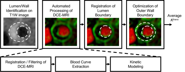
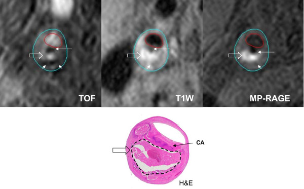
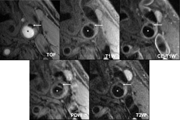
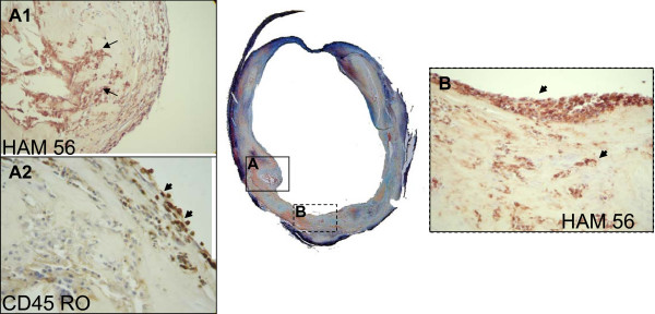
Atherosclerosis is a chronic, progressive, inflammatory disease affecting many vascular beds. Disease progression leads to acute cardiovascular events such as myocardial infarction, stroke and death. The diseased carotid alone is responsible for one third of the 700,000 new or recurrent strokes occurring yearly in the United States. Imaging plays an important role in the management of atherosclerosis, and cardiovascular magnetic resonance (CMR) of the carotid vessel wall is one promising modality in the evaluation of patients with carotid atherosclerotic disease. Advances in carotid vessel wall CMR allow comprehensive assessment of morphology inside the wall, contributing substantial disease-specific information beyond luminal stenosis. Although carotid vessel wall CMR has not been widely used to screen for carotid atherosclerotic disease, many trials support its potential for this indication. This review summarizes the current state of knowledge regarding carotid vessel wall CMR and its potential clinical application for management of carotid atherosclerotic disease.

摘要

动脉粥样硬化是一种影响许多血管床的慢性、进行性、炎症性疾病。疾病的进展导致急性心血管事件,如心肌梗死、中风和死亡。仅病变颈动脉就导致了美国每年发生的 70 万例新的或复发的中风中的三分之一。影像学在动脉粥样硬化的管理中起着重要作用,颈动脉血管壁的心血管磁共振(CMR)是评估颈动脉粥样硬化疾病患者的一种很有前途的方式。颈动脉血管壁 CMR 的进步允许对血管壁内部的形态进行全面评估,提供了除管腔狭窄以外的大量特定于疾病的信息。尽管颈动脉血管壁 CMR 尚未广泛用于筛查颈动脉粥样硬化疾病,但许多试验支持其在该适应证中的应用潜力。这篇综述总结了目前关于颈动脉血管壁 CMR 的知识状态及其在颈动脉粥样硬化疾病管理中的潜在临床应用。

https://cdn.ncbi.nlm.nih.gov/pmc/blobs/4f22/2806867/312f3d4cd0ee/1532-429X-11-53-8.jpg
https://cdn.ncbi.nlm.nih.gov/pmc/blobs/4f22/2806867/34e9518f1909/1532-429X-11-53-1.jpg
https://cdn.ncbi.nlm.nih.gov/pmc/blobs/4f22/2806867/384d2002f4d7/1532-429X-11-53-2.jpg
https://cdn.ncbi.nlm.nih.gov/pmc/blobs/4f22/2806867/5c0fea7463d4/1532-429X-11-53-3.jpg
https://cdn.ncbi.nlm.nih.gov/pmc/blobs/4f22/2806867/97c5beffa161/1532-429X-11-53-4.jpg
https://cdn.ncbi.nlm.nih.gov/pmc/blobs/4f22/2806867/c7092eb22ab7/1532-429X-11-53-5.jpg
https://cdn.ncbi.nlm.nih.gov/pmc/blobs/4f22/2806867/b8a513d099d4/1532-429X-11-53-6.jpg
https://cdn.ncbi.nlm.nih.gov/pmc/blobs/4f22/2806867/2277a1472172/1532-429X-11-53-7.jpg
https://cdn.ncbi.nlm.nih.gov/pmc/blobs/4f22/2806867/312f3d4cd0ee/1532-429X-11-53-8.jpg

相似文献

1
Cardiovascular magnetic resonance in carotid atherosclerotic disease.颈动脉粥样硬化性疾病的心血管磁共振。
J Cardiovasc Magn Reson. 2009 Dec 15;11(1):53. doi: 10.1186/1532-429X-11-53.
2
Co-existing intracranial and extracranial carotid artery atherosclerotic plaques and recurrent stroke risk: a three-dimensional multicontrast cardiovascular magnetic resonance study.颅内和颅外颈动脉粥样硬化斑块并存与复发性卒中风险:一项三维多对比心血管磁共振研究
J Cardiovasc Magn Reson. 2016 Dec 2;18(1):90. doi: 10.1186/s12968-016-0309-3.
3
Magnetic [corrected] resonance imaging [corrected] features of the disruption-prone and the disrupted carotid plaque.易破裂和已破裂颈动脉斑块的磁共振成像特征[已修正]
JACC Cardiovasc Imaging. 2009 Jul;2(7):883-96. doi: 10.1016/j.jcmg.2009.03.013.
4
Determinants of carotid atherosclerotic plaque burden in a stroke-free population.无卒中人群中颈动脉粥样硬化斑块负荷的决定因素。
Atherosclerosis. 2016 Dec;255:186-192. doi: 10.1016/j.atherosclerosis.2016.10.030. Epub 2016 Oct 14.
5
Multi-contrast atherosclerosis characterization (MATCH) of carotid plaque with a single 5-min scan: technical development and clinical feasibility.单次5分钟扫描对颈动脉斑块进行多对比动脉粥样硬化特征分析(MATCH):技术发展与临床可行性
J Cardiovasc Magn Reson. 2014 Jul 25;16(1):53. doi: 10.1186/s12968-014-0053-5.
6
Atherosclerotic plaque imaging: the last frontier for cardiac magnetic resonance.动脉粥样硬化斑块成像:心脏磁共振成像的最后前沿领域。
JACC Cardiovasc Imaging. 2009 Jul;2(7):916-8. doi: 10.1016/j.jcmg.2009.05.002.
7
High resolution 3D diffusion cardiovascular magnetic resonance of carotid vessel wall to detect lipid core without contrast media.无需造影剂的高分辨率三维扩散心血管磁共振成像用于检测颈动脉血管壁脂质核。
J Cardiovasc Magn Reson. 2014 Sep 17;16(1):67. doi: 10.1186/s12968-014-0067-z.
8
In-vivo quantitative T2 mapping of carotid arteries in atherosclerotic patients: segmentation and T2 measurement of plaque components.动脉粥样硬化患者颈动脉活体定量 T2 成像:斑块成分的分割和 T2 值测量。
J Cardiovasc Magn Reson. 2013 Aug 16;15(1):69. doi: 10.1186/1532-429X-15-69.
9
Expansive arterial remodeling of the carotid arteries and its effect on atherosclerotic plaque composition and vulnerability: an in-vivo black-blood 3T CMR study in symptomatic stroke patients.颈动脉的扩张性动脉重塑及其对动脉粥样硬化斑块成分和易损性的影响:一项针对有症状中风患者的体内黑血3T磁共振成像研究
J Cardiovasc Magn Reson. 2016 Mar 3;18:11. doi: 10.1186/s12968-016-0229-2.
10
Quantification of Lipid-Rich Core in Carotid Atherosclerosis Using Magnetic Resonance T Mapping: Relation to Clinical Presentation.利用磁共振T成像技术对颈动脉粥样硬化中富含脂质的核心进行定量分析:与临床表现的关系
JACC Cardiovasc Imaging. 2017 Jul;10(7):747-756. doi: 10.1016/j.jcmg.2016.06.013. Epub 2016 Oct 12.

引用本文的文献

1
Visualization of Atherosclerotic Plaques Paired with Joheksol 350 (Omnipaque).动脉粥样硬化斑块与碘海醇350(欧乃派克)联合成像。
Biomedicines. 2025 Feb 7;13(2):399. doi: 10.3390/biomedicines13020399.
2
High-Risk Plaque Features in the Non-stenosing Carotid Artery, How Frequently is This Reported? A Retrospective Study.非狭窄性颈动脉的高危斑块特征,其报告频率如何?一项回顾性研究。
Neurohospitalist. 2024 Sep 14:19418744241283858. doi: 10.1177/19418744241283858.
3
The role of non-stenosing carotid artery plaques in embolic stroke of undetermined source, is it a silent offender? A review of literature.

本文引用的文献

1
Arterial remodeling in [corrected] subclinical carotid artery disease.[已纠正]亚临床颈动脉疾病中的动脉重塑。
JACC Cardiovasc Imaging. 2009 Dec;2(12):1381-9. doi: 10.1016/j.jcmg.2009.08.007.
2
Tyrosine polyethylene glycol (PEG)-micelle magnetic resonance contrast agent for the detection of lipid rich areas in atherosclerotic plaque.用于检测动脉粥样硬化斑块中富含脂质区域的酪氨酸聚乙二醇(PEG)胶束磁共振对比剂。
Magn Reson Med. 2009 Nov;62(5):1195-201. doi: 10.1002/mrm.22103.
3
Geometric and compositional appearance of atheroma in an angiographically normal carotid artery in patients with atherosclerosis.
非狭窄颈动脉斑块在不明来源栓塞性卒中中的作用,它是沉默的罪犯吗?文献复习。
Interv Neuroradiol. 2024 Oct;30(5):759-767. doi: 10.1177/15910199221143172. Epub 2022 Nov 30.
4
Automated segmentation of the individual branches of the carotid arteries in contrast-enhanced MR angiography using DeepMedic.使用 DeepMedic 对对比增强磁共振血管造影中的颈动脉各分支进行自动分割。
BMC Med Imaging. 2021 Feb 27;21(1):38. doi: 10.1186/s12880-021-00568-6.
5
Carotid Plaque Fibrous Cap Thickness Measurement by ARFI Variance of Acceleration: In Vivo Human Results.基于声辐射力脉冲(ARFI)加速度方差的颈动脉斑块纤维帽厚度测量:在体人体研究结果。
IEEE Trans Med Imaging. 2020 Dec;39(12):4383-4390. doi: 10.1109/TMI.2020.3019184. Epub 2020 Nov 30.
6
Navigator-based reacquisition and estimation of motion-corrupted data: Application to multi-echo spin echo for carotid wall MRI.基于导航器的运动 corrupted 数据的重新采集与估计:在颈动脉壁 MRI 的多回波自旋回波中的应用。 (注:原文中“motion-corrupted”可能有误,推测可能是“motion-corrupted”,直译为“运动损坏的”,这里意译为“运动干扰的”可能更合适,但按照要求未修改原文翻译)
Magn Reson Med. 2020 Jun;83(6):2026-2041. doi: 10.1002/mrm.28063. Epub 2019 Nov 7.
7
Four Different Carotid Atherosclerotic Behaviors Based on Luminal Stenosis and Plaque Characteristics in Symptomatic Patients: An in Vivo Study.基于有症状患者管腔狭窄和斑块特征的四种不同颈动脉粥样硬化行为:一项体内研究
Diagnostics (Basel). 2019 Oct 2;9(4):137. doi: 10.3390/diagnostics9040137.
8
Hypertriglyceridemic Waist: A Simple Marker of High-Risk Atherosclerosis Features Associated With Excess Visceral Adiposity/Ectopic Fat.高甘油三酯性腰围:一种与内脏肥胖/异位脂肪相关的高风险动脉粥样硬化特征的简单标志物。
J Am Heart Assoc. 2018 Apr 13;7(8):e008139. doi: 10.1161/JAHA.117.008139.
9
First in-human radiation dosimetry of Ga-NODAGA-RGDyK.镓-氮杂环辛四乙酸-环肽-RGDyK的首次人体辐射剂量测定。
EJNMMI Res. 2017 Dec;7(1):43. doi: 10.1186/s13550-017-0288-x. Epub 2017 May 18.
10
Comparison of Gated and Ungated Black-Blood Fast Spin-echo MRI of Carotid Vessel Wall at 3T.3T下颈动脉血管壁门控与非门控黑血快速自旋回波MRI的比较
Magn Reson Med Sci. 2016 Jul 11;15(3):266-72. doi: 10.2463/mrms.mp.2014-0133. Epub 2015 Nov 6.
动脉粥样硬化患者血管造影正常的颈动脉中的动脉粥样硬化的几何和组成外观。
AJNR Am J Neuroradiol. 2010 Feb;31(2):311-6. doi: 10.3174/ajnr.A1793. Epub 2009 Sep 24.
4
Reproducibility of fibrous cap status assessment of carotid artery plaques by contrast-enhanced MRI.通过对比增强磁共振成像评估颈动脉斑块纤维帽状态的可重复性。
Stroke. 2009 Sep;40(9):3017-21. doi: 10.1161/STROKEAHA.109.555052. Epub 2009 Jun 25.
5
Utility of high resolution MR imaging to assess carotid plaque morphology: a comparison of acute symptomatic, recently symptomatic and asymptomatic patients with carotid artery disease.高分辨率磁共振成像在评估颈动脉斑块形态学中的应用:急性症状性、近期症状性和无症状性颈动脉疾病患者的比较。
Atherosclerosis. 2009 Dec;207(2):434-9. doi: 10.1016/j.atherosclerosis.2009.05.002. Epub 2009 May 13.
6
Moderate carotid artery stenosis: MR imaging-depicted intraplaque hemorrhage predicts risk of cerebrovascular ischemic events in asymptomatic men.中度颈动脉狭窄:磁共振成像显示的斑块内出血可预测无症状男性发生脑血管缺血事件的风险。
Radiology. 2009 Aug;252(2):502-8. doi: 10.1148/radiol.2522080792. Epub 2009 Jun 9.
7
The ATHEROMA (Atorvastatin Therapy: Effects on Reduction of Macrophage Activity) Study. Evaluation using ultrasmall superparamagnetic iron oxide-enhanced magnetic resonance imaging in carotid disease.动脉粥样硬化(阿托伐他汀治疗:对巨噬细胞活性降低的影响)研究。使用超小型超顺磁性氧化铁增强磁共振成像对颈动脉疾病进行评估。
J Am Coll Cardiol. 2009 Jun 2;53(22):2039-50. doi: 10.1016/j.jacc.2009.03.018.
8
Macrophage-specific lipid-based nanoparticles improve cardiac magnetic resonance detection and characterization of human atherosclerosis.巨噬细胞特异性脂质纳米颗粒改善了人类动脉粥样硬化的心脏磁共振检测与特征分析。
JACC Cardiovasc Imaging. 2009 May;2(5):637-47. doi: 10.1016/j.jcmg.2008.08.009.
9
Cardiovascular magnetic resonance parameters of atherosclerotic plaque burden improve discrimination of prior major adverse cardiovascular events.动脉粥样硬化斑块负荷的心血管磁共振参数可改善对既往主要不良心血管事件的辨别能力。
J Cardiovasc Magn Reson. 2009 Apr 24;11(1):10. doi: 10.1186/1532-429X-11-10.
10
Temporal dependence of in vivo USPIO-enhanced MRI signal changes in human carotid atheromatous plaques.体内超顺磁性氧化铁增强磁共振成像信号变化在人类颈动脉粥样硬化斑块中的时间依赖性
Neuroradiology. 2009 Jul;51(7):457-65. doi: 10.1007/s00234-009-0523-x. Epub 2009 Mar 20.