Institute of Membrane and Systems Biology, University of Leeds, Leeds, United Kingdom.
PLoS One. 2009 Dec 14;4(12):e8312. doi: 10.1371/journal.pone.0008312.
Many ion channels are preferentially located in caveolae where compartmentalisation/scaffolding with signal transduction components regulates their activity. Channels that are mechanosensitive may be additionally dependent on caveolar control of the mechanical state of the membrane. Here we test which mechanism underlies caveolar-regulation of the mechanosensitive I(Cl,swell) channel in the adult cardiac myocyte.
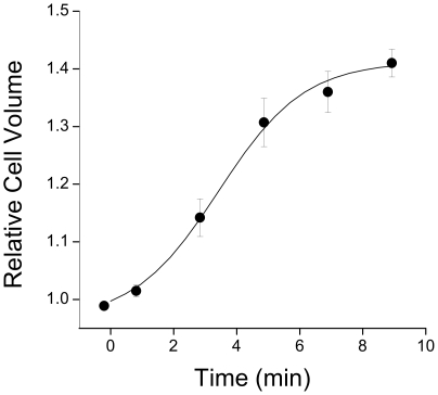
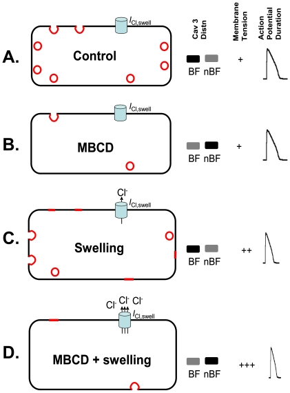
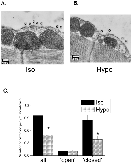
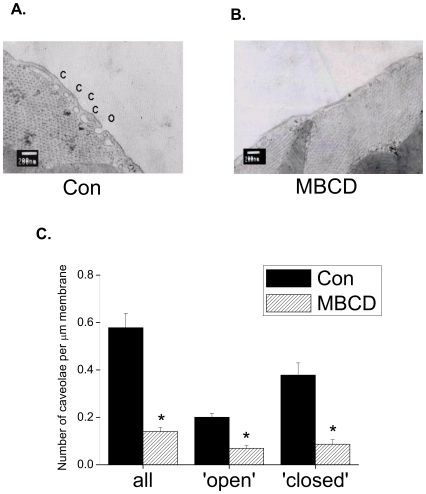
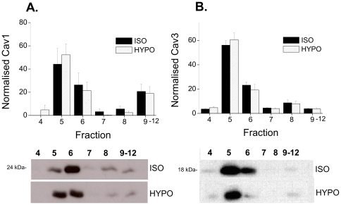
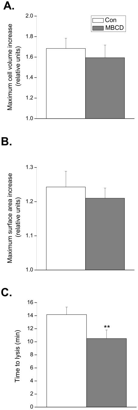
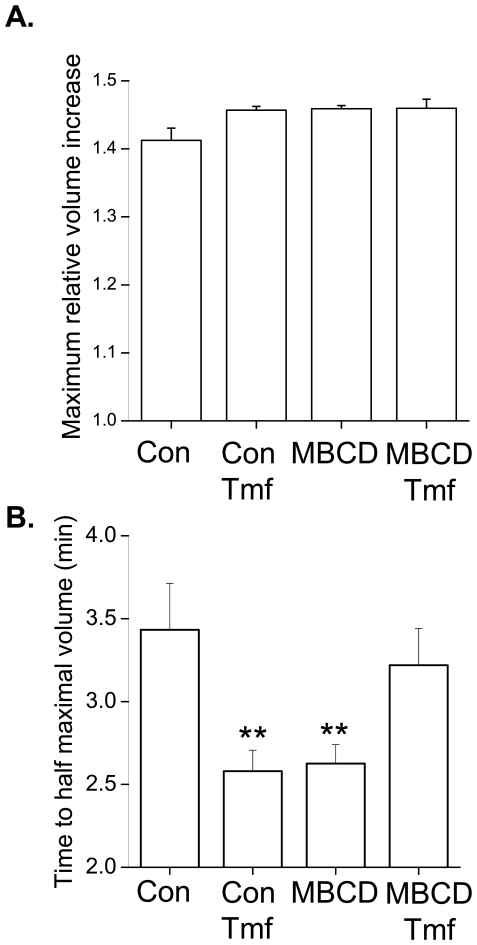
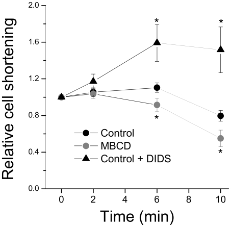
METHODOLOGY/PRINCIPAL FINDINGS: Rat ventricular myocytes were exposed to solution of 0.02 tonicity (T; until lysis), 0.64T for 10-15 min (swelling), and/or methyl-beta-cyclodextrin (MBCD; to disrupt caveolae). MBCD and 0.64T swelling reduced the number of caveolae visualised by electron microscopy by 75 and 50% respectively. MBCD stimulated translocation of caveolin 3 from caveolae-enriched buoyant membrane fractions, but both caveolin 1 and 3 remained in buoyant fractions after swelling. I(Cl,swell) inhibition in control cells decreased time to half-maximal volume (t(0.5,vol); 0.64T), consistent with a role for I(Cl,swell) in volume regulation. MBCD-treated cells showed reduced time to lysis (0.02T) and t(0.5,vol) (0.64T) compared with controls. The negative inotropic response to swelling (an index of I(Cl,swell) activation) was enhanced by MBCD.
CONCLUSIONS/SIGNIFICANCE: These data show that disrupting caveolae removes essential membrane reserves, which speeds swelling in hyposmotic conditions, and thereby promotes activation of I(Cl,swell). They illustrate a general principle whereby caveolae as a membrane reserve limit increases in membrane tension during stretch/swelling thereby restricting mechanosensitive channel activation.
许多离子通道优先定位于质膜凹陷( caveolae )中,在质膜凹陷中,信号转导组分的区室化/支架作用调节其活性。机械敏感性通道可能还依赖于质膜机械状态的质膜凹陷控制。在此,我们测试机械敏感性 I(Cl,swell) 通道在成年心肌细胞中的质膜凹陷调节的机制。
方法/主要发现:将大鼠心室肌细胞暴露于 0.02 渗透压( T ;直至裂解)、0.64T 10-15min(肿胀)和/或甲基-β-环糊精( MBCD ;破坏质膜凹陷)的溶液中。MBCD 和 0.64T 肿胀分别使电子显微镜下观察到的质膜凹陷数量减少 75%和 50%。MBCD 刺激 caveolin 3 从富含质膜凹陷的浮力膜部分易位,但肿胀后 caveolin 1 和 3 仍保留在浮力部分。在对照细胞中,I(Cl,swell) 抑制使达到半最大体积的时间( t(0.5,vol) ; 0.64T )缩短,表明 I(Cl,swell) 在体积调节中起作用。与对照相比,MBCD 处理的细胞在低渗条件下的裂解时间( 0.02T )和 t(0.5,vol) ( 0.64T )均缩短。肿胀引起的负性变力反应( I(Cl,swell) 激活的指标)被 MBCD 增强。
结论/意义:这些数据表明,破坏质膜凹陷会去除必需的膜储备,这会加速低渗条件下的肿胀,从而促进 I(Cl,swell) 的激活。它们说明了一个普遍的原理,即质膜凹陷作为膜储备限制了拉伸/肿胀过程中膜张力的增加,从而限制了机械敏感性通道的激活。