Neurologia Clinica, Università Campus Bio-Medico di Roma, Italy.
BMC Neurosci. 2009 Dec 20;10:151. doi: 10.1186/1471-2202-10-151.
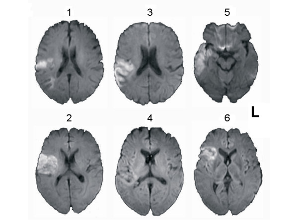
By mapping the dynamics of brain reorganization, functional magnetic resonance imaging MRI (fMRI) has allowed for significant progress in understanding cerebral plasticity phenomena after a stroke. However, cerebro-vascular diseases can affect blood oxygen level dependent (BOLD) signal. Cerebral autoregulation is a primary function of cerebral hemodynamics, which allows to maintain a relatively constant blood flow despite changes in arterial blood pressure and perfusion pressure. Cerebral autoregulation is reported to become less effective in the early phases post-stroke. This study investigated whether any impairment of cerebral hemodynamics that occurs during the acute and the subacute phases of ischemic stroke is related to changes in BOLD response. We enrolled six aphasic patients affected by acute stroke. All patients underwent a Transcranial Doppler to assess cerebral autoregulation (Mx index) and fMRI to evaluate the amplitude and the peak latency (time to peak-TTP) of BOLD response in the acute (i.e., within four days of stroke occurrence) and the subacute (i.e., between five and twelve days after stroke onset) stroke phases.
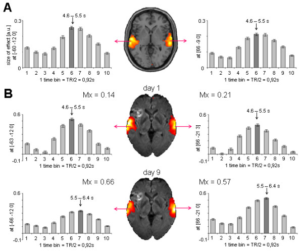
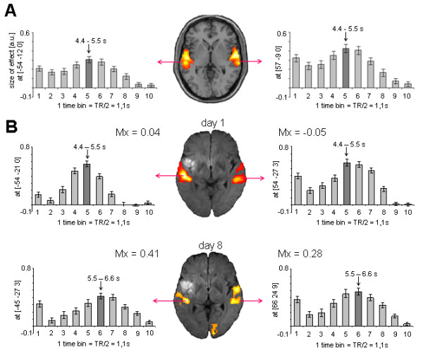
As patients advanced from the acute to subacute stroke phase, the affected hemisphere presented a BOLD TTP increase (p = 0.04) and a deterioration of cerebral autoregulation (Mx index increase, p = 0.046). A similar but not significant trend was observed also in the unaffected hemisphere. When the two hemispheres were grouped together, BOLD TTP delay was significantly related to worsening cerebral autoregulation (Mx index increase) (Spearman's rho = 0.734; p = 0.01).
The hemodynamic response function subtending BOLD signal may present a delay in peak latency that arises as patients advance from the acute to the subacute stroke phase. This delay is related to the deterioration of cerebral hemodynamics. These findings suggest that remodeling the fMRI hemodynamic response function in the different phases of stroke may optimize the detection of BOLD signal changes.
通过绘制大脑重组的动态图,功能磁共振成像(fMRI)在理解中风后大脑可塑性现象方面取得了重大进展。然而,脑血管疾病会影响血氧水平依赖(BOLD)信号。脑自动调节是脑血流动力学的主要功能,它可以在动脉血压和灌注压变化时保持相对稳定的血流量。据报道,脑自动调节在中风后早期阶段的效果会降低。本研究旨在探讨中风急性期和亚急性期发生的任何脑血流动力学损伤是否与 BOLD 反应的变化有关。我们纳入了 6 名患有急性中风的失语症患者。所有患者均接受经颅多普勒超声检查以评估脑自动调节(Mx 指数),并进行 fMRI 检查以评估急性(即中风发生后 4 天内)和亚急性期(即中风发生后 5 至 12 天)BOLD 反应的振幅和峰值时程(达峰时间-TTP)。
随着患者从急性期向亚急性期进展,受影响的半球 BOLD TTP 增加(p=0.04),脑自动调节恶化(Mx 指数增加,p=0.046)。在未受影响的半球也观察到类似但不显著的趋势。当将两个半球归为一组时,BOLD TTP 延迟与脑自动调节恶化(Mx 指数增加)显著相关(Spearman 相关系数=0.734;p=0.01)。
在患者从急性期向亚急性期进展时,支撑 BOLD 信号的血流动力学反应函数可能会出现峰值时程的延迟。这种延迟与脑血流动力学的恶化有关。这些发现表明,在中风的不同阶段重塑 fMRI 血流动力学反应功能可能会优化 BOLD 信号变化的检测。