Zuccarello Biagio, Spada Antonella, Turiaco Nunzio, Villari Daniela, Parisi Saveria, Francica Isabella, Fazzari Carmine, Pederiva Federica, Tovar Juan A
Policlinico Universitario G.Martino, 98125 Messina, Italy.
Int J Pediatr. 2009;2009:695837. doi: 10.1155/2009/695837. Epub 2009 Jul 27.
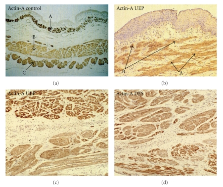
Introduction and Aim. Disorders of esophageal motility causing dysphagia and gastroesophageal reflux are frequent in survivors to esophageal atresia (EA) and distal tracheoesophageal fistula (TEF). The aim of the present study was to investigate the histologic and immunohistochemical features in both esophageal atretic segments to further understand the nature of the motor disorders observed in these patients. Material and Methods. Esophageal specimens from 12 newborns with EA/TEF and 5 newborns dead of unrelated causes were examined. The specimens were fixed in 5% buffered formalin, included in paraffin and cut in 5 micron sections that were stained with hematoxilin and eosin (H and E), and immunohistochemical stainings for Actin, S-100 protein, Neurofilament, Neuron-Specific-Enolase, Chromogranin A and Peripherin were evaluated under the microscope. Results. In controls, the distribution of the neural elements was rather homogenous at both levels of the esophagus. In contrast, the atretic segments showed quantitative and qualitative differences between them with sparser nervous tissue in the distal one in comparison with the proximal one and with controls. Conclusions. These results further support the assumption that histomorphological alterations of the muscular and nervous elements within the esophageal wall might contribute to esophageal dysmotility in patients surviving neonatal operations for EA/TEF.
引言与目的。食管闭锁(EA)和远端气管食管瘘(TEF)幸存者中,导致吞咽困难和胃食管反流的食管动力障碍很常见。本研究的目的是调查食管闭锁段的组织学和免疫组化特征,以进一步了解这些患者中观察到的运动障碍的本质。材料与方法。检查了12例患有EA/TEF的新生儿和5例因无关原因死亡的新生儿的食管标本。标本用5%缓冲甲醛固定,包埋在石蜡中,切成5微米厚的切片,用苏木精和伊红(H&E)染色,并在显微镜下评估肌动蛋白、S-100蛋白、神经丝、神经元特异性烯醇化酶、嗜铬粒蛋白A和外周蛋白的免疫组化染色。结果。在对照组中,食管两个水平的神经成分分布相当均匀。相比之下,闭锁段之间在数量和质量上存在差异,远端闭锁段的神经组织比近端闭锁段和对照组更稀疏。结论。这些结果进一步支持了这样一种假设,即食管壁内肌肉和神经成分的组织形态学改变可能导致EA/TEF新生儿手术后存活患者的食管运动障碍。