Department of Radiology, The Research Institute of Radiological Science, Yonsei University College of Medicine, Seoul, Korea.
Yonsei Med J. 2009 Dec 31;50(6):838-44. doi: 10.3349/ymj.2009.50.6.838. Epub 2009 Dec 18.
To evaluate the value of breast MRI in analysis of papillomas of the breast.
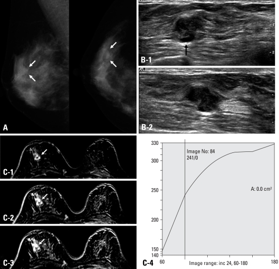
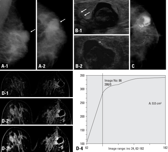
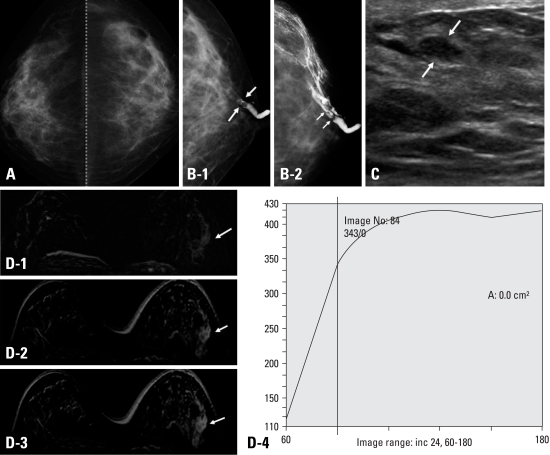
From 1996 to 2004, 94 patients underwent surgery due to papillomas of the breast. Among them, 21 patients underwent 3D fast low angle shot (FLASH) dynamic breast MRI. Eight masses were palpable and 11 of 21 patients had nipple discharge. Two radiologists indifferently analyzed the location, size of the lesions and shape, margin of the masses, multiplicity and ductal relation. The MRI findings were categorized according to breast imaging reporting and data system (BI-RADS) lexicon. The amount and pattern of enhancement and associated findings were also evaluated according to BI-RADS. We then compared the MRI findings with galactography, mammography and breast ultrasonography (US) and examined histopathologic correlation.
On breast MRI, the lesion size was 0.4-1.59 cm, and 18 patients showed subareolar location. On 4.25 cm (mean 1.54) dynamic enhanced images, imaging findings showed mass (n = 10), intracystic mass (n = 3), focus (n = 5), ductal enhancement (n = 2), and segmental enhancement (n = 1). In cases of the masses, the shapes of the masses were round (n = 4), lobulated (n = 3), and irregular (n = 6), and margins were circumscribed (n = 6), microlobulated (n = 5), and indistinct (n = 2). The enhancement patterns were homogeneous enhancement (n = 7), heterogeneous (n = 3) or rim enhancement (n = 3).
The contrast enhanced dynamic breast MRI was highly sensitive for diagnosis of breast papillomas. MRI could play a key role in the pre-operative work-up for multiple papillomas and papillomatosis.
评估乳腺 MRI 在乳腺纤维瘤分析中的价值。
1996 年至 2004 年间,94 例因乳腺纤维瘤而行手术治疗的患者,其中 21 例行 3D 快速低角采集(FLASH)动态乳腺 MRI 检查。8 个肿块可触及,21 例中有 11 例乳头溢液。两位放射科医生分别分析病变的位置、大小、形状、边缘、多发性和导管关系。MRI 表现根据乳腺影像报告和数据系统(BI-RADS)词汇进行分类。根据 BI-RADS,还评估了强化的程度和模式以及相关表现。然后,我们将 MRI 结果与乳管造影、乳房 X 线摄影和乳房超声(US)进行比较,并检查组织病理学相关性。
乳腺 MRI 上病变大小为 0.4-1.59cm,18 例位于乳晕下。在 4.25cm(平均 1.54cm)的动态增强图像上,影像学表现为肿块(n=10)、囊内肿块(n=3)、局灶性病变(n=5)、导管强化(n=2)和节段性强化(n=1)。在肿块中,肿块形状为圆形(n=4)、分叶状(n=3)和不规则形(n=6),边缘为边界清楚(n=6)、微分叶状(n=5)和模糊(n=2)。强化模式为均匀强化(n=7)、不均匀强化(n=3)或边缘强化(n=3)。
对比增强动态乳腺 MRI 对乳腺纤维瘤的诊断具有很高的敏感性。MRI 在多发性纤维瘤和纤维瘤病的术前评估中可能发挥关键作用。