• 文献检索
  • 文档翻译
  • 深度研究
  • 学术资讯
  • Suppr Zotero 插件Zotero 插件
  • 邀请有礼
  • 套餐&价格
  • 历史记录
应用&插件
Suppr Zotero 插件Zotero 插件浏览器插件Mac 客户端Windows 客户端微信小程序
定价
高级版会员购买积分包购买API积分包
服务
文献检索文档翻译深度研究API 文档MCP 服务
关于我们
关于 Suppr公司介绍联系我们用户协议隐私条款
关注我们

Suppr 超能文献

核心技术专利:CN118964589B侵权必究
粤ICP备2023148730 号-1Suppr @ 2026

文献检索

告别复杂PubMed语法,用中文像聊天一样搜索,搜遍4000万医学文献。AI智能推荐,让科研检索更轻松。

立即免费搜索

文件翻译

保留排版,准确专业,支持PDF/Word/PPT等文件格式,支持 12+语言互译。

免费翻译文档

深度研究

AI帮你快速写综述,25分钟生成高质量综述,智能提取关键信息,辅助科研写作。

立即免费体验

影像学测量足后弓:长轴位片比足后弓位片更可靠。

Measuring hindfoot alignment radiographically: the long axial view is more reliable than the hindfoot alignment view.

机构信息

Department of Orthopaedic Surgery, Academic Medical Center, Amsterdam, The Netherlands.

出版信息

Skeletal Radiol. 2010 Nov;39(11):1103-8. doi: 10.1007/s00256-009-0857-9. Epub 2010 Jan 9.

DOI:10.1007/s00256-009-0857-9
PMID:20062985
原文链接:https://pmc.ncbi.nlm.nih.gov/articles/PMC2939352/
Abstract

BACKGROUND

Hindfoot malalignment is a recognized cause of foot and ankle disability. For preoperative planning and clinical follow-up, reliable radiographic assessment of hindfoot alignment is important. The long axial radiographic view and the hindfoot alignment view are commonly used for this purpose. However, their comparative reliabilities are unknown. As hindfoot varus or valgus malalignment is most pronounced during mid-stance of gait, a unilateral weight-bearing stance, in comparison with a bilateral stance, could increase measurement reliability. The purpose of this study was to compare the intra- and interobserver reliability of hindfoot alignment measurements of both radiographic views in bilateral and unilateral stance.

MATERIALS AND METHODS

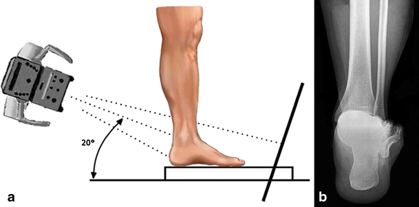
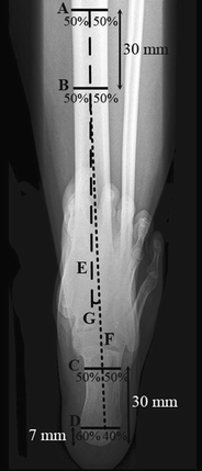
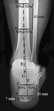
A hindfoot alignment view and a long axial view were acquired from 18 healthy volunteers in bilateral and unilateral weight-bearing stances. Hindfoot alignment was defined as the angular deviation between the tibial anatomical axis and the calcaneus longitudinal axis from the radiographs. Repeat measurements of hindfoot alignment were performed by nine orthopaedic examiners.

RESULTS

Measurements from the hindfoot alignment view gave intra- and interclass correlation coefficients (CCs) of 0.72 and 0.58, respectively, for bilateral stance and 0.91 and 0.49, respectively, for unilateral stance. The long axial view showed, respectively, intra- and interclass CCs of 0.93 and 0.79 for bilateral stance and 0.91 and 0.58 for unilateral stance.

CONCLUSION

The long axial view is more reliable than the hindfoot alignment view or the angular measurement of hindfoot alignment. Although intra-observer reliability is good/excellent for both methods, only the long axial view leads to good interobserver reliability. A unilateral weight-bearing stance does not lead to greater reliability of measurement.

摘要

背景

后足对线不良是足部和踝关节功能障碍的公认原因。为了术前规划和临床随访,可靠的后足对线放射学评估非常重要。长轴位和后足对线位常用于此目的。然而,它们的比较可靠性尚不清楚。由于后足内翻或外翻对线不良在步态的中间支撑期最为明显,与双侧支撑相比,单侧负重支撑可能会增加测量的可靠性。本研究的目的是比较双侧和单侧负重支撑时两种影像学视图后足对线测量的内部和观察者间可靠性。

材料和方法

从 18 名健康志愿者的双侧和单侧负重支撑中获得后足对线位和长轴位。后足对线定义为从影像学图像上胫骨解剖轴和跟骨长轴之间的角度偏差。由 9 名矫形检查者重复进行后足对线测量。

结果

后足对线位测量的组内和组间相关系数(CC)分别为双侧支撑时 0.72 和 0.58,单侧支撑时 0.91 和 0.49。长轴位分别显示双侧支撑时的组内和组间 CC 为 0.93 和 0.79,单侧支撑时为 0.91 和 0.58。

结论

长轴位比后足对线位或后足对线角度测量更可靠。虽然两种方法的观察者内可靠性都很好/优秀,但只有长轴位才能产生良好的观察者间可靠性。单侧负重支撑不会导致测量的可靠性更高。

https://cdn.ncbi.nlm.nih.gov/pmc/blobs/19ce/2939352/83f23a23b0ba/256_2009_857_Fig4_HTML.jpg
https://cdn.ncbi.nlm.nih.gov/pmc/blobs/19ce/2939352/e0b26fc05512/256_2009_857_Fig1_HTML.jpg
https://cdn.ncbi.nlm.nih.gov/pmc/blobs/19ce/2939352/50cc6b527c0a/256_2009_857_Fig2_HTML.jpg
https://cdn.ncbi.nlm.nih.gov/pmc/blobs/19ce/2939352/0fe86378a426/256_2009_857_Fig3_HTML.jpg
https://cdn.ncbi.nlm.nih.gov/pmc/blobs/19ce/2939352/83f23a23b0ba/256_2009_857_Fig4_HTML.jpg

相似文献

1
Measuring hindfoot alignment radiographically: the long axial view is more reliable than the hindfoot alignment view.影像学测量足后弓:长轴位片比足后弓位片更可靠。
Skeletal Radiol. 2010 Nov;39(11):1103-8. doi: 10.1007/s00256-009-0857-9. Epub 2010 Jan 9.
2
Radiologic analysis of hindfoot alignment: Comparison of Méary, long axial, and hindfoot alignment views.后足对线的放射学分析:梅里位、长轴位和后足对线位视图的比较。
Orthop Traumatol Surg Res. 2017 Dec;103(8):1211-1216. doi: 10.1016/j.otsr.2017.08.014. Epub 2017 Sep 28.
3
Does the subtalar joint compensate for ankle malalignment in end-stage ankle arthritis?距下关节是否能代偿终末期踝关节关节炎中的踝关节对线不良?
Clin Orthop Relat Res. 2015 Jan;473(1):318-25. doi: 10.1007/s11999-014-3960-8. Epub 2014 Oct 15.
4
Correlation of Clinical Evaluation and Radiographic Hindfoot Alignment in Stage II Adult-Acquired Flatfoot Deformity.II期成人获得性平足畸形的临床评估与影像学后足对线的相关性
Foot Ankle Int. 2018 Jul;39(7):771-779. doi: 10.1177/1071100718762113. Epub 2018 Mar 28.
5
Intraoperative assessment of hindfoot alignment using C-arm fluoroscopy.术中使用 C 臂透视评估后足对线。
J Orthop Sci. 2024 Sep;29(5):1248-1254. doi: 10.1016/j.jos.2023.08.014. Epub 2023 Oct 13.
6
Hindfoot coronal alignment: a modified radiographic method.后足冠状面排列:一种改良的放射学方法。
Foot Ankle Int. 1999 Dec;20(12):818-25. doi: 10.1177/107110079902001212.
7
Hindfoot alignment measurements: rotation-stability of measurement techniques on hindfoot alignment view and long axial view radiographs.足后弓对线测量:足后弓对线视图和长轴视图 X 射线片测量技术的旋转稳定性。
AJR Am J Roentgenol. 2011 Sep;197(3):578-82. doi: 10.2214/AJR.10.5728.
8
Republication of "How Accurate Is Clinical Evaluation in Hindfoot Coronal Alignment?".《后足冠状面排列的临床评估有多准确?》再版
Foot Ankle Orthop. 2023 Jul 23;8(3):24730114231188111. doi: 10.1177/24730114231188111. eCollection 2023 Jul.
9
Three-dimensional hindfoot alignment measurements based on biplanar radiographs: comparison with standard radiographic measurements.基于双平面 X 线片的三维后足对线测量:与标准 X 线测量的比较。
Skeletal Radiol. 2013 Apr;42(4):493-8. doi: 10.1007/s00256-012-1543-x. Epub 2012 Nov 20.
10
Diagnostic performance of MRI measurements to assess hindfoot malalignment. An assessment of four measurement techniques.MRI 测量评估后足对线不良的诊断性能。四种测量技术的评估。
Eur Radiol. 2013 Sep;23(9):2594-601. doi: 10.1007/s00330-013-2839-5. Epub 2013 Apr 12.

引用本文的文献

1
Correlation between varus-type knee osteoarthritis severity and hindfoot alignment: Analysis of radiographs in the long-leg weight-bearing anteroposterior view.膝内翻型膝骨关节炎严重程度与后足对线的相关性:长腿负重前后位X线片分析
PLoS One. 2025 Jun 3;20(6):e0324974. doi: 10.1371/journal.pone.0324974. eCollection 2025.
2
Can Calcaneofibular Ligament Repair Correct Hindfoot Varus Alignment in the Management of Chronic Lateral Ankle Instability?在慢性外侧踝关节不稳的治疗中,跟腓韧带修复能否纠正后足内翻对线?
Cureus. 2025 Apr 22;17(4):e82768. doi: 10.7759/cureus.82768. eCollection 2025 Apr.
3
Impact of subtalar joint debridement on fusion rates and outcomes in tibiotalocalcaneal arthrodesis.

本文引用的文献

1
Interrater reliability of subtalar neutral, calcaneal inversion and eversion.跟骨中立位、内翻/外翻位的测量者间信度。
J Orthop Sports Phys Ther. 1990;12(1):10-5. doi: 10.2519/jospt.1990.12.1.10.
2
Correlation of compensatory alignment of the subtalar joint to the progression of primary osteoarthritis of the ankle.距下关节代偿性对线与踝关节原发性骨关节炎进展的相关性。
Foot Ankle Int. 2008 Apr;29(4):400-6. doi: 10.3113/FAI.2008.0400.
3
[Osteotomies of the distal tibia and hindfoot for ankle realignment].[胫骨远端和后足截骨术用于踝关节重新对线]
距下关节清创术对胫距跟关节融合率及疗效的影响。
J Orthop Surg Res. 2025 Apr 23;20(1):408. doi: 10.1186/s13018-025-05817-w.
4
Does Timing of Hindfoot Reconstruction Before or After Primary Total Knee Arthroplasty Impact Reoperation Rates?后足重建在初次全膝关节置换术前或术后进行的时机是否会影响再次手术率?
Foot Ankle Orthop. 2025 Mar 17;10(1):24730114251318735. doi: 10.1177/24730114251318735. eCollection 2025 Jan.
5
The association between varus knee deformity and morphological changes in the foot and ankle in patients with end-stage varus knee osteoarthritis.终末期膝内翻型骨关节炎患者膝内翻畸形与足踝形态学改变之间的关联
J Orthop Surg Res. 2025 Jan 7;20(1):15. doi: 10.1186/s13018-024-05433-0.
6
Coxa pedis: can calcaneal pronation angle be considered a predictive sign of medial plantar arch overload?足跟内翻角能否作为内侧纵弓过载的预测指标?
Radiol Med. 2024 Jun;129(6):925-933. doi: 10.1007/s11547-024-01815-w. Epub 2024 Apr 24.
7
Assessment of Changes in Hindfoot Alignment of Total Ankle Arthroplasty for Ankle Osteoarthritis on Weightbearing Subtalar X-ray View.负重下距下关节X线片评估踝关节骨关节炎全踝关节置换术后后足对线的变化
Foot Ankle Orthop. 2023 Oct 28;8(4):24730114231205299. doi: 10.1177/24730114231205299. eCollection 2023 Oct.
8
TIBIOCALCANEAL ARTHRODESIS: A COMPARISON OF ANTERIOR APPROACH AND TRANSFIBULAR APPROACH.胫跟关节融合术:前路与经腓骨入路的比较
Acta Ortop Bras. 2023 Oct 23;31(5):e267148. doi: 10.1590/1413-785220233105e267148. eCollection 2023.
9
Modified hindfoot alignment radiological evaluation and application in the assessment of flatfoot.改良后足对线放射学评估及其在扁平足评估中的应用。
BMC Musculoskelet Disord. 2023 Aug 29;24(1):683. doi: 10.1186/s12891-023-06824-w.
10
Republication of "How Accurate Is Clinical Evaluation in Hindfoot Coronal Alignment?".《后足冠状面排列的临床评估有多准确?》再版
Foot Ankle Orthop. 2023 Jul 23;8(3):24730114231188111. doi: 10.1177/24730114231188111. eCollection 2023 Jul.
Orthopade. 2008 Mar;37(3):212-8, 220-3. doi: 10.1007/s00132-008-1214-8.
4
Long leg calcaneal axial and hindfoot alignment radiographic views for frontal plane assessment.用于额状面评估的长腿跟骨轴位和后足对线X线片。
J Am Podiatr Med Assoc. 2008 Jan-Feb;98(1):75-8. doi: 10.7547/0980075.
5
Low tibial osteotomy for varus-type osteoarthritis of the ankle.低位胫骨截骨术治疗踝关节内翻型骨关节炎
J Bone Joint Surg Br. 2006 Jul;88(7):909-13. doi: 10.1302/0301-620X.88B7.17325.
6
Double calcaneal osteotomy: realignment considerations in eight patients.双跟骨截骨术:8例患者的重新排列考量
J Am Podiatr Med Assoc. 2005 Jan-Feb;95(1):53-9. doi: 10.7547/0950053.
7
Static rearfoot alignment: a comparison of clinical and radiographic measures.静态后足对线:临床测量与影像学测量的比较
J Am Podiatr Med Assoc. 2005 Jan-Feb;95(1):26-33. doi: 10.7547/0950026.
8
A systematic approach to evaluation of the rearfoot, ankle, and leg in reconstructive surgery.重建手术中后足、踝关节及小腿评估的系统方法。
J Am Podiatr Med Assoc. 2005 Jan-Feb;95(1):2-12. doi: 10.7547/0950002.
9
Measuring alignment of the hindfoot.测量后足的对线情况。
J Biomech Eng. 2004 Jun;126(3):357-62. doi: 10.1115/1.1762897.
10
Radiographic assessment of the hindfoot and ankle.后足和踝关节的影像学评估。
Clin Podiatr Med Surg. 2004 Jul;21(3):295-304, v. doi: 10.1016/j.cpm.2004.03.004.