Nicholas Cory R, Haston Kelly M, Pera Renee A Reijo
Institute for Stem Cell Biology and Regenerative Medicine, Department of Obstetrics and Gynecology, Stanford University, Palo Alto, CA, USA.
BMC Dev Biol. 2010 Jan 8;10:2. doi: 10.1186/1471-213X-10-2.
Female reproductive potential, or the ability to propagate life, is limited in mammals with the majority of oocytes lost before birth. In mice, surviving perinatal oocytes are enclosed in ovarian follicles for subsequent oocyte development and function in the adult. Before birth, fetal germ cells of both sexes develop in clusters, or germline cysts, in the undifferentiated gonad. Upon sex determination of the fetal gonad, germ cell cysts become organized into testicular or ovarian cord-like structures and begin to interact with gonadal somatic cells. Although germline cysts and testicular cords are required for spermatogenesis, the role of cyst and ovarian cord formation in mammalian oocyte development and female fertility has not been determined.
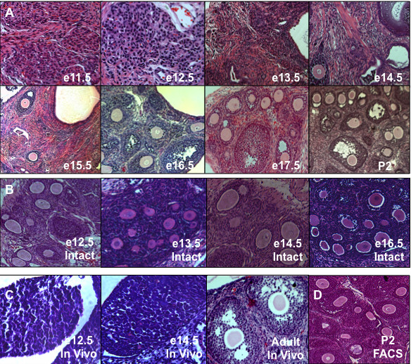
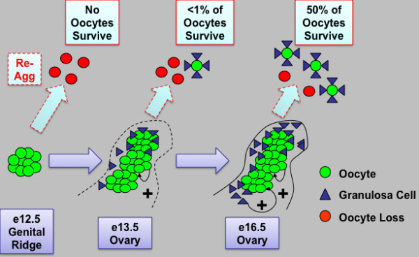
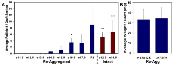
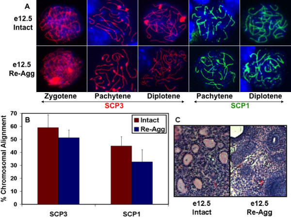
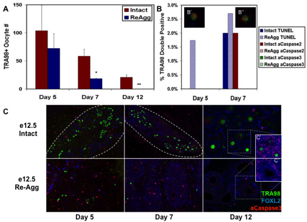
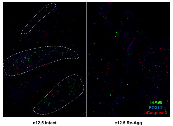
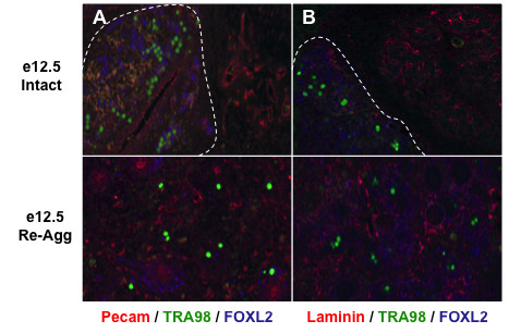
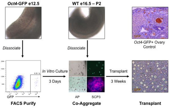
Here, we examine whether intact fetal ovarian germ and somatic cell cord structures are required for oocyte development using mouse gonad re-aggregation and transplantation to disrupt gonadal organization. We observed that germ cells from disrupted female gonad prior to embryonic day e13.5 completed prophase I of meiosis but did not survive following transplantation. Furthermore, re-aggregated ovaries from e13.5 to e15.5 developed with a reduced number of oocytes. Oocyte loss occurred before follicle formation and was associated with an absence of ovarian cord structure and ovary disorganization. However, disrupted ovaries from e16.5 or later were resistant to the re-aggregation impairment and supported robust oocyte survival and development in follicles.
Thus, we demonstrate a critical window of oocyte development from e13.5 to e16.5 in the intact fetal mouse ovary, corresponding to the establishment of ovarian cord structure, which promotes oocyte interaction with neighboring ovarian somatic granulosa cells before birth and imparts oocytes with competence to survive and develop in follicles. Because germline cyst and ovarian cord structures are conserved in the human fetal ovary, the identification of genetic components and molecular mechanisms of pre-follicle stage germ and somatic cell structures may be important for understanding human female infertility. In addition, this work provides a foundation for development of a robust fetal ovarian niche and transplantation based system to direct stem cell-derived oocyte differentiation as a potential therapeutic strategy for the treatment of infertility.
雌性生殖潜能,即繁衍生命的能力,在哺乳动物中是有限的,大多数卵母细胞在出生前就已丢失。在小鼠中,围产期存活的卵母细胞被包裹在卵巢卵泡中,以供成年后卵母细胞的后续发育和发挥功能。出生前,两性的胎儿生殖细胞在未分化的性腺中以细胞团或种系囊肿的形式发育。在胎儿性腺性别确定后,生殖细胞囊肿会组织成睾丸或卵巢索状结构,并开始与性腺体细胞相互作用。虽然种系囊肿和睾丸索对于精子发生是必需的,但囊肿和卵巢索形成在哺乳动物卵母细胞发育和雌性生育能力中的作用尚未确定。
在此,我们利用小鼠性腺重新聚集和移植来破坏性腺组织,研究完整的胎儿卵巢生殖细胞和体细胞索状结构对于卵母细胞发育是否必要。我们观察到,在胚胎第13.5天(e13.5)之前破坏的雌性性腺中的生殖细胞完成了减数分裂前期I,但移植后未能存活。此外,从e13.5到e15.5重新聚集的卵巢发育出的卵母细胞数量减少。卵母细胞丢失发生在卵泡形成之前,并且与卵巢索结构缺失和卵巢组织紊乱有关。然而,来自e16.5或更晚时期的破坏卵巢对重新聚集损伤具有抗性,并支持卵泡中卵母细胞的强劲存活和发育。
因此,我们证明了在完整的胎儿小鼠卵巢中,从e13.5到e16.5是卵母细胞发育的关键时期,这与卵巢索结构的建立相对应,卵巢索结构在出生前促进卵母细胞与相邻的卵巢颗粒体细胞相互作用,并赋予卵母细胞在卵泡中存活和发育的能力。由于人类胎儿卵巢中种系囊肿和卵巢索结构是保守的,鉴定卵泡前阶段生殖细胞和体细胞结构的遗传成分和分子机制可能对理解人类女性不孕症很重要。此外,这项工作为开发强大的胎儿卵巢微环境和基于移植的系统奠定了基础,以指导干细胞衍生的卵母细胞分化,作为治疗不孕症的潜在治疗策略。