Laboratory for Atherosclerosis and Metabolic Research, University of California Davis Medical Center, Sacramento, California, USA.
Diabetes Care. 2010 Apr;33(4):861-8. doi: 10.2337/dc09-1799. Epub 2010 Jan 12.
Individuals with type 2 diabetes have a myriad of metabolic aberrations including increased inflammation, increasing their cardiovascular risk. Toll-like receptors (TLRs) and their ligands play a key role in insulin resistance and atherosclerosis. However, there is a paucity of data examining the expression and activity of TLRs in type 2 diabetes. Thus, in the present study, we examined TLR2 and TLR4 mRNA and protein expression, their ligands, and signaling in monocytes of recently diagnosed type 2 diabetic patients.
TLR mRNA, protein expression, TLR ligands, and TLR signaling were measured in freshly isolated monocytes from healthy human control subjects (n = 23) and type 2 diabetic subjects (n = 23) using real-time RT-PCR, Western blot, and flow cytometric assays.
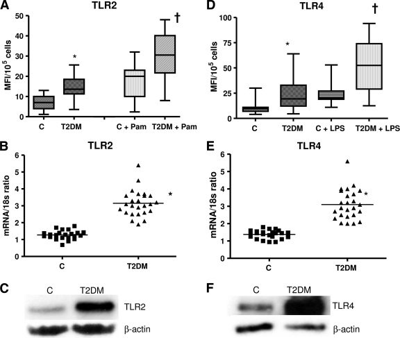
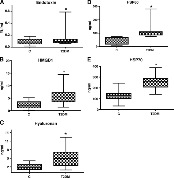
Type 2 diabetic subjects had significantly increased TLR2, TLR4 mRNA, and protein in monocytes compared with control subjects (P < 0.05). Increased TLR2 and TLR4 expression correlated with BMI, homeostasis model assessment-insulin resistance (HOMA-IR), glucose, A1C, N(epsilon)-(carboxymethyl) lysine (CML), and free fatty acid (FFA). Ligands of TLR2 and TLR4, namely, HSP60, HSP70, HMGB1, endotoxin, and hyaluronan levels, were elevated in type 2 diabetic subjects and positively correlated with TLR2 and TLR4. Type 2 diabetic subjects showed increased MyD88, phosphorylated IRAK-1, Trif, TICAM-1, IRF-3, and NF-kappaB p65 expression in monocytes compared with control subjects. Furthermore, TLR-MyD88-NF-kappaB signaling resulted in elevated levels of cytokines (P < 0.05), but increased interleukin (IL)-1beta, interferon (IFN)-gamma, and endotoxin were not significant when adjusted for BMI.
In this comprehensive study, we make the novel observation that TLR2 and TLR4 expression and their ligands, signaling, and functional activation are increased in recently diagnosed type 2 diabetes and contribute to the proinflammatory state.
2 型糖尿病患者存在多种代谢异常,包括炎症增加,从而增加其心血管风险。Toll 样受体 (TLR) 及其配体在胰岛素抵抗和动脉粥样硬化中发挥关键作用。然而,目前关于 2 型糖尿病中 TLR 的表达和活性的数据很少。因此,在本研究中,我们检测了新诊断的 2 型糖尿病患者单核细胞中 TLR2 和 TLR4 的 mRNA 和蛋白表达、其配体和信号转导。
使用实时 RT-PCR、Western blot 和流式细胞术检测来自健康对照者(n=23)和 2 型糖尿病患者(n=23)的新鲜分离单核细胞中的 TLR mRNA、蛋白表达、TLR 配体和 TLR 信号。
与对照组相比,2 型糖尿病患者的单核细胞中 TLR2、TLR4 mRNA 和蛋白显著增加(P<0.05)。TLR2 和 TLR4 表达的增加与 BMI、稳态模型评估-胰岛素抵抗 (HOMA-IR)、血糖、A1C、N(epsilon)-(羧甲基)赖氨酸 (CML) 和游离脂肪酸 (FFA) 相关。TLR2 和 TLR4 的配体,即热休克蛋白 60(HSP60)、热休克蛋白 70(HSP70)、高迁移率族蛋白 B1(HMGB1)、内毒素和透明质酸水平,在 2 型糖尿病患者中升高,并与 TLR2 和 TLR4 呈正相关。与对照组相比,2 型糖尿病患者的单核细胞中 MyD88、磷酸化 IRAK-1、Trif、TICAM-1、IRF-3 和 NF-κB p65 表达增加。此外,TLR-MyD88-NF-κB 信号导致细胞因子水平升高(P<0.05),但调整 BMI 后,IL-1β、IFN-γ 和内毒素的增加并不显著。
在这项全面的研究中,我们首次观察到 TLR2 和 TLR4 的表达及其配体、信号转导和功能激活在新诊断的 2 型糖尿病中增加,并导致促炎状态。