State Key Laboratory of Reproductive Biology, Institute of Zoology, Chinese Academy of Sciences, Beijing, PR China.
Reprod Biol Endocrinol. 2010 Jan 15;8:5. doi: 10.1186/1477-7827-8-5.
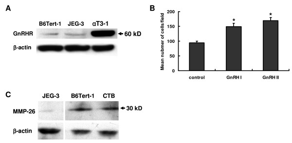
Matrix metalloproteinase-26 (MMP-26), one of the main mediators of extracellular matrix (ECM) degradation, has been shown to exist in trophoblasts of human placenta and to play a role in trophoblast cell invasion. However, little is known about the regulation of MMP-26 expression in human trophoblasts. Recently, gonadotropin-releasing hormone I (GnRH I) and GnRH II have been shown to regulate the expression of MMP-2, MMP-9/tissue inhibitor of metalloproteinases 1 (TIMP-1), and urokinase plasminogen activator (uPA)/plasminogen activator inhibitor (PAI) in human trophoblasts, suggesting that these two hormones may work as paracrine and/or autocrine regulators in modulating the activities of various protease systems at the feto-maternal interface. In this study, we determined the regulatory effects of GnRH I and GnRH II on the expression of MMP-26 in human immortalized cytotrophoblast-like cell line, B6Tert-1.
Real-time PCR was used to quantify mRNA levels of MMP-26 in human trophoblast-like cell line, B6Tert-1 and primary cultured cytotrophoblasts. Western blotting was used to characterize the expression of MMP-26 and the phosphorylation of c-Jun NH2-terminal kinase (JNK) and extracellular signal-regulated kinase 1/2 (ERK1/2) in B6Tert-1 cells after treatment with GnRH I and GnRH II.
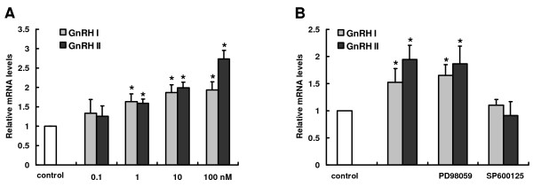
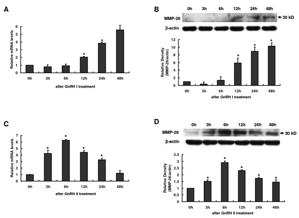
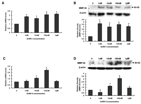
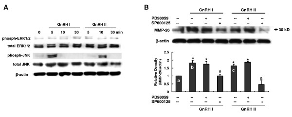
We found that GnRH I increased MMP-26 expression in B6Tert-1 cells after 12 h of treatment at both the mRNA and protein level, while GnRH II increased MMP-26 expression beginning at 3 h of treatment. Treatment of GnRH I at 1 nM resulted in maximal increase of MMP-26 mRNA and protein levels, whereas GnRH II treatment at a concentration of 100 nM was required to induce maximal increase in MMP-26 expression. In addition, we demonstrated that the activation of JNK, but not ERK1/2, was required for GnRH I and II-stimulated MMP-26 production in B6Tert-1 cells and primary cytotrophoblasts.
These novel findings indicated that GnRH I and II could up-regulate MMP-26 expression through the JNK signaling pathway in human trophoblast-like/trophoblast cells.
基质金属蛋白酶-26(MMP-26)是细胞外基质(ECM)降解的主要介质之一,已在人胎盘滋养细胞中存在,并在滋养细胞浸润中发挥作用。然而,关于人滋养细胞中 MMP-26 表达的调控知之甚少。最近,促性腺激素释放激素 I(GnRH I)和 GnRH II 已被证明可调节人滋养细胞中 MMP-2、MMP-9/金属蛋白酶组织抑制剂 1(TIMP-1)和尿激酶纤溶酶原激活物(uPA)/纤溶酶原激活物抑制剂(PAI)的表达,表明这两种激素可能作为旁分泌和/或自分泌调节剂,在调节胎-母界面各种蛋白酶系统的活性方面发挥作用。在这项研究中,我们确定了 GnRH I 和 GnRH II 对人永生化绒毛细胞样细胞系 B6Tert-1 中 MMP-26 表达的调节作用。
实时 PCR 用于定量人滋养细胞样细胞系 B6Tert-1 和原代培养的绒毛细胞中 MMP-26 的 mRNA 水平。Western blot 用于表征 GnRH I 和 GnRH II 处理后 B6Tert-1 细胞中 MMP-26 的表达以及 c-Jun NH2-末端激酶(JNK)和细胞外信号调节激酶 1/2(ERK1/2)的磷酸化。
我们发现 GnRH I 在处理 12 小时后可增加 B6Tert-1 细胞中 MMP-26 的 mRNA 和蛋白水平,而 GnRH II 在处理 3 小时后即可增加 MMP-26 的表达。1 nM GnRH I 处理可导致 MMP-26 mRNA 和蛋白水平的最大增加,而 100 nM GnRH II 处理可诱导 MMP-26 表达的最大增加。此外,我们证明 JNK 的激活,但不是 ERK1/2 的激活,是 GnRH I 和 II 刺激 B6Tert-1 细胞和原代绒毛细胞中 MMP-26 产生所必需的。
这些新发现表明,GnRH I 和 II 可以通过人绒毛细胞样/滋养细胞中的 JNK 信号通路上调 MMP-26 的表达。