Department of Medicine (Cardiovascular Division and the Center for Cardiovascular Research), Washington University School of Medicine, St. Louis, MO, USA.
Cardiovasc Pathol. 2010 Nov-Dec;19(6):e233-40. doi: 10.1016/j.carpath.2009.12.002. Epub 2010 Jan 25.
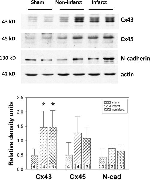
We have recently shown that native murine ventricular fibroblasts express both connexin43 (Cx43) and Cx45, and that the level of Cx43 expression influences intercellular coupling and cell proliferation. Relatively little is known, however, about how myocardial infarction (MI) influences expression of Cx43, or how altered Cx43 expression may affect fibroblast function post-MI. Fibroblasts are critical for infarct healing and post-infarct ventricular remodeling. They can couple electrically with cardiac myocytes and influence myocardial activation patterns. Thus, Cx43 remodeling and the level of intercellular communication in fibroblasts expressed in the infarcted heart were the subject of the present investigation.
Fibroblasts were isolated from both infarct scar and remote, noninfarcted regions of murine hearts 6 d after coronary ligation. Expression levels of Cx43, α-smooth muscle actin and N-cadherin were quantified by immunoblotting. Gap junctional intercellular communication was quantified by Lucifer yellow dye transfer.
Fibroblasts isolated from infarcted hearts exhibited marked up-regulation of Cx43 protein expression and enhanced intercellular coupling. Exogenous administration of transforming growth factor-β (TGF-β) to fibroblast cultures from normal, non-operated hearts produced comparable up-regulation of Cx43, suggesting that increased intercellular communication between fibroblasts in infarct and peri-infarct regions may be secondary to activation of a TGF-β pathway. Unlike cardiac myocytes that down-regulate Cx43, presumably to limit intercellular transmission of biochemical mediators of ischemic injury, fibroblasts may up-regulate Cx43 to maintain electrical and metabolic coupling at a time when intercellular communication is compromised.
我们最近发现,天然的鼠心室成纤维细胞表达连接蛋白 43(Cx43)和 Cx45,并且 Cx43 的表达水平影响细胞间偶联和细胞增殖。然而,关于心肌梗死(MI)如何影响 Cx43 的表达,或者改变的 Cx43 表达如何影响 MI 后成纤维细胞的功能,我们知之甚少。成纤维细胞对于梗死愈合和 MI 后心室重构至关重要。它们可以与心肌细胞电偶联并影响心肌激活模式。因此,Cx43 重构和梗死心脏中成纤维细胞的细胞间通讯水平是本研究的主题。
在冠状动脉结扎后 6 天,从 MI 疤痕和远离 MI 的非梗死区的鼠心中分离成纤维细胞。通过免疫印迹定量测定 Cx43、α-平滑肌肌动蛋白和 N-钙黏蛋白的表达水平。通过 Lucifer yellow 染料转移定量测定缝隙连接细胞间通讯。
从 MI 心脏中分离的成纤维细胞表现出 Cx43 蛋白表达的显著上调和增强的细胞间偶联。外源性给予 TGF-β转化生长因子-β(TGF-β)到来自正常、未手术的心脏的成纤维细胞培养物中,产生了类似的 Cx43 上调,这表明梗死和梗死周围区域成纤维细胞之间增加的细胞间通讯可能是 TGF-β 途径激活的结果。与下调 Cx43 以限制缺血性损伤生化介质的细胞间传递的心肌细胞不同,成纤维细胞可能上调 Cx43 以在细胞间通讯受损时维持电和代谢偶联。