Department of Internal Medicine, School of Medicine, Federal University of Rio de Janeiro, Clementino Fraga Filho University Hospital, Rua Prof. Rodolpho Paulo Rocco, 255, Rio de Janeiro, 21941-913, Brasil.
BMC Vet Res. 2010 Jan 29;6:6. doi: 10.1186/1746-6148-6-6.
Domestic dogs and cats are very well known to develop chronic hepatic diseases, including hepatic lipidosis and cirrhosis. Ultrasonographic examination is extensively used to detect them. However, there are still few reports on the use of the ultrasound B-mode scan in correlation with histological findings to evaluate diffuse hepatic changes in rodents, which represent the most important animal group used in experimental models of liver diseases. The purpose of this study was to determine the reliability of ultrasound findings in the assessment of fatty liver disease and cirrhosis when compared to histological results in Wistar rats by following up a murine model of chronic hepatic disease.
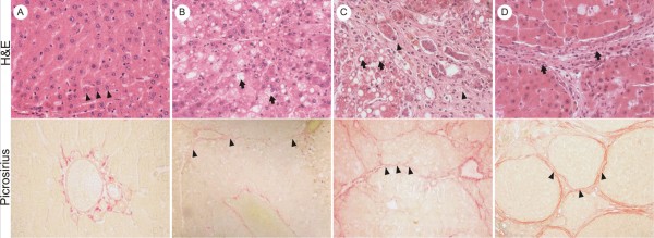
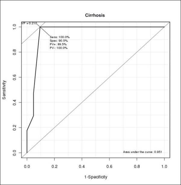
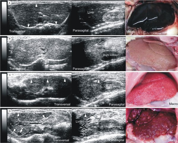
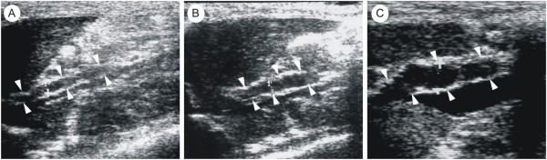
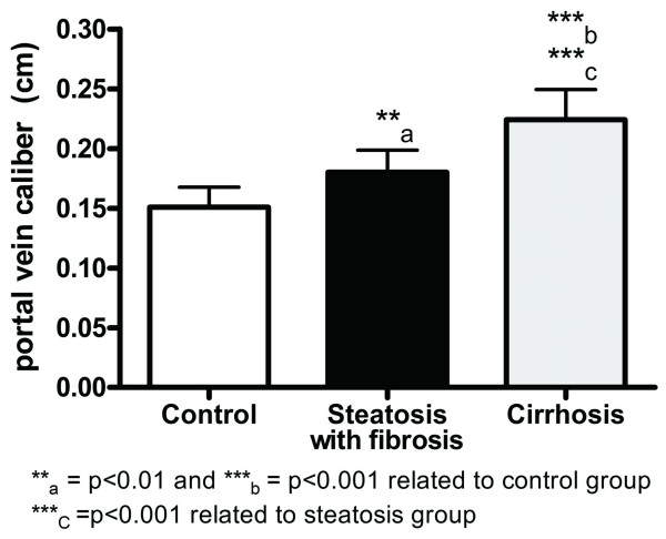
Forty Wistar rats (30 treated, 10 controls) were included. Liver injury was induced by dual exposure to CCl4 and ethanol for 4, 8 and 15 weeks. Liver echogenicity, its correlation to the right renal cortex echogenicity, measurement of portal vein diameter (PVD) and the presence of ascites were evaluated and compared to histological findings of hepatic steatosis and cirrhosis. Liver echogenicity correlated to hepatic steatosis when it was greater or equal to the right renal cortex echogenicity, with a sensitivity of 90%, specificity of 100%, positive and negative predictive values of 100% and 76.9% respectively, and accuracy of 92.5%. Findings of heterogeneous liver echogenicity and irregular surface correlated to liver cirrhosis with a sensitivity of 70.6%, specificity of 100%, positive and negative predictive values of 100% and 82.1% respectively, and accuracy of 87.5%. PVD was significantly increased in both steatotic and cirrhotic rats; however, the later had greater diameters. PVD cut-off point separating steatosis from cirrhosis was 2.1 mm (sensitivity of 100% and specificity of 90.5%). One third of cirrhotic rats presented with ascites.
The use of ultrasound imaging in the follow-up of murine diffuse liver disease models is feasible and efficient, especially when the studied parameters are used in combination. The potential implication of this study is to provide a non-invasive method that allows follow-up studies of fatty liver disease and cirrhosis of individual rats for pre-clinical drug or cell based therapies.
家犬和家猫众所周知会发生慢性肝病,包括肝脂肪变性和肝硬化。超声检查广泛用于检测这些疾病。然而,在评估啮齿动物弥漫性肝改变方面,与组织学结果相关的超声 B 型扫描在代表用于肝脏疾病实验模型的最重要动物群体中使用的报道仍然很少。本研究的目的是通过跟踪慢性肝病的小鼠模型,确定超声检查在评估 Wistar 大鼠脂肪肝和肝硬化方面的可靠性,与组织学结果相比。
纳入 40 只 Wistar 大鼠(30 只治疗组,10 只对照组)。肝损伤由 CCl4 和乙醇双重暴露 4、8 和 15 周引起。评估和比较肝脏回声、与右肾皮质回声的相关性、门静脉直径(PVD)的测量以及腹水的存在,以评估肝脂肪变性和肝硬化的组织学结果。当肝脏回声大于或等于右肾皮质回声时,与肝脂肪变性相关,其灵敏度为 90%,特异性为 100%,阳性和阴性预测值分别为 100%和 76.9%,准确性为 92.5%。不均匀的肝脏回声和不规则表面的发现与肝硬化相关,其灵敏度为 70.6%,特异性为 100%,阳性和阴性预测值分别为 100%和 82.1%,准确性为 87.5%。PVD 在脂肪变性和肝硬化大鼠中均显著增加;然而,后者的直径更大。将 PVD 截断值从 2.1mm 分开,以区分脂肪变性和肝硬化,其灵敏度为 100%,特异性为 90.5%。三分之一的肝硬化大鼠出现腹水。
在慢性肝疾病模型的监测中使用超声成像既可行又有效,特别是当研究参数联合使用时。本研究的潜在意义是提供一种非侵入性方法,可以对个体大鼠的脂肪肝和肝硬化进行随访,用于临床前药物或细胞治疗。