Department of Surgery, University Hospital, De Pintelaan 185, Ghent, Belgium.
Br J Cancer. 2010 Mar 2;102(5):837-43. doi: 10.1038/sj.bjc.6605535. Epub 2010 Feb 2.
Recently, low-molecular-weight heparins (LMWHs) were found to confer a survival advantage in cancer patients. The mechanism underlying this observation is unclear, but may involve inhibition of tumour angiogenesis. We aimed to examine the effects of nadroparin on tumour angiogenesis using a dorsal skinfold window chamber model in the Syrian hamster.
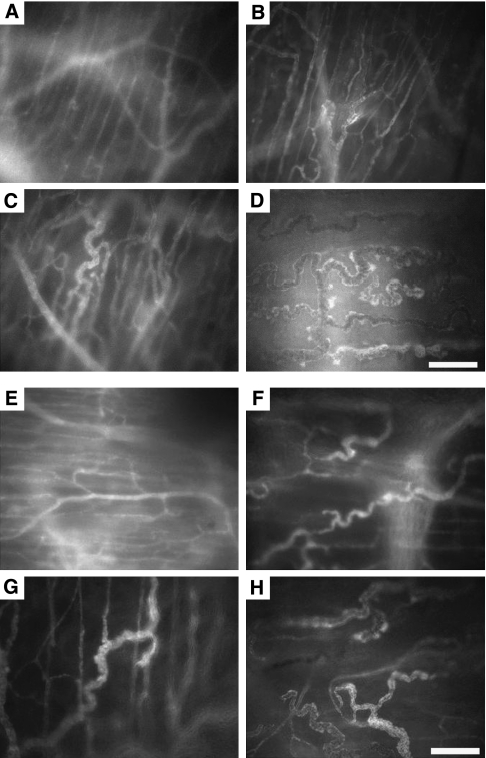
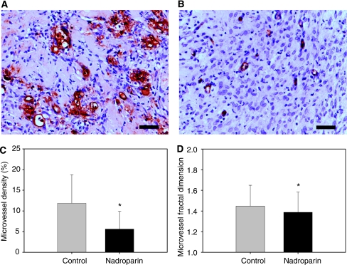
AMel-3 and HAP-T1 tumours were grown in donor animals and fragments implanted in the window chambers. Animals (N=46) were treated with 200 IU of nadroparin or saline for 10 days. Repeated intravital fluorescence microscopy was performed to calculate functional microcirculatory parameters: number (N) and length (L) of microvessels, vascular area fraction (AF), and red blood cell velocity (V). Microvessel density (MVD), fractal dimension, and pericyte coverage were assessed histologically.
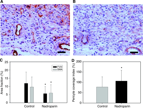
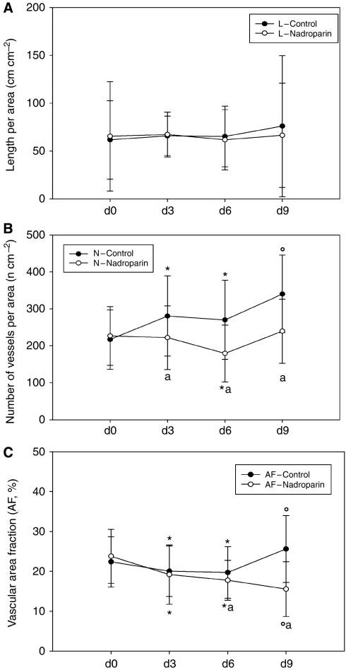
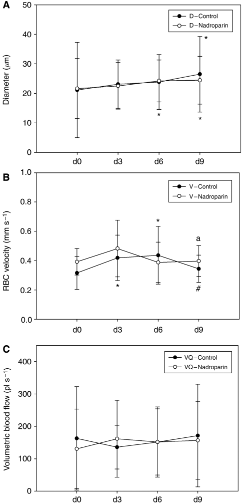
Active angiogenesis was observed in control animals, resulting in a significant increase in N, L, and AF. In nadroparin-treated animals, however, N and L did not increase whereas AF decreased significantly. Both groups showed an initial increase in V, but nadroparin treatment resulted in an earlier decrease in red blood cell velocity over time. Compared with control animals, nadroparin-treated animals showed a significantly lower MVD and fractal dimension but significantly higher pericyte coverage index (PCI).
Taken together, these results suggest that the LMWH nadroparin inhibits tumour angiogenesis and results in microvessel normalisation.
最近发现低分子肝素(LMWHs)可使癌症患者的生存获益。这种观察结果的机制尚不清楚,但可能涉及抑制肿瘤血管生成。我们旨在使用叙利亚仓鼠背部皮肤窗室模型来检查那屈肝素对肿瘤血管生成的影响。
在供体动物中生长 AMel-3 和 HAP-T1 肿瘤,并将肿瘤碎片植入窗室。将动物(N=46)用 200 IU 的那屈肝素或生理盐水治疗 10 天。重复进行活体荧光显微镜检查以计算功能微循环参数:微血管数量(N)和长度(L)、血管面积分数(AF)和红细胞速度(V)。通过组织学评估微血管密度(MVD)、分形维数和周细胞覆盖率。
在对照组动物中观察到活跃的血管生成,导致 N、L 和 AF 显著增加。然而,在那屈肝素治疗的动物中,N 和 L 没有增加,而 AF 显著降低。两组动物的 V 均先增加,但那屈肝素治疗后红细胞速度随时间的早期下降。与对照组动物相比,那屈肝素治疗组的 MVD 和分形维数明显降低,但周细胞覆盖率指数(PCI)明显升高。
综上所述,这些结果表明 LMWH 那屈肝素抑制肿瘤血管生成并导致微血管正常化。