• 文献检索
  • 文档翻译
  • 深度研究
  • 学术资讯
  • Suppr Zotero 插件Zotero 插件
  • 邀请有礼
  • 套餐&价格
  • 历史记录
应用&插件
Suppr Zotero 插件Zotero 插件浏览器插件Mac 客户端Windows 客户端微信小程序
定价
高级版会员购买积分包购买API积分包
服务
文献检索文档翻译深度研究API 文档MCP 服务
关于我们
关于 Suppr公司介绍联系我们用户协议隐私条款
关注我们

Suppr 超能文献

核心技术专利:CN118964589B侵权必究
粤ICP备2023148730 号-1Suppr @ 2026

文献检索

告别复杂PubMed语法,用中文像聊天一样搜索,搜遍4000万医学文献。AI智能推荐,让科研检索更轻松。

立即免费搜索

文件翻译

保留排版,准确专业,支持PDF/Word/PPT等文件格式,支持 12+语言互译。

免费翻译文档

深度研究

AI帮你快速写综述,25分钟生成高质量综述,智能提取关键信息,辅助科研写作。

立即免费体验

踝关节的软骨骨缺损:为何会疼痛?

Osteochondral defects in the ankle: why painful?

机构信息

Department of Orthopaedic Surgery, Academic Medical Center, University of Amsterdam, PO Box 22660, 1100 DD Amsterdam, The Netherlands.

出版信息

Knee Surg Sports Traumatol Arthrosc. 2010 May;18(5):570-80. doi: 10.1007/s00167-010-1064-x. Epub 2010 Feb 12.

DOI:10.1007/s00167-010-1064-x
PMID:20151110
原文链接:https://pmc.ncbi.nlm.nih.gov/articles/PMC2855020/
Abstract

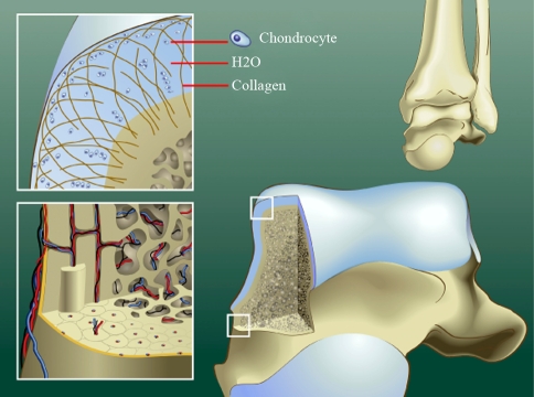
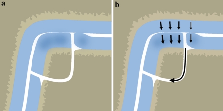
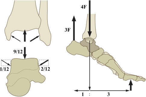
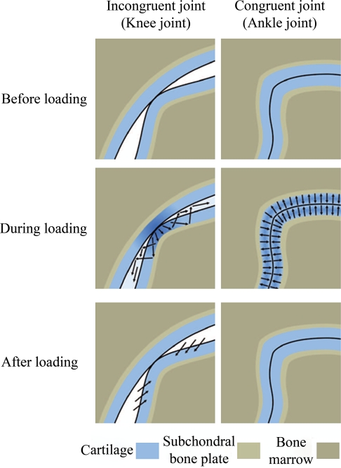
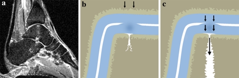
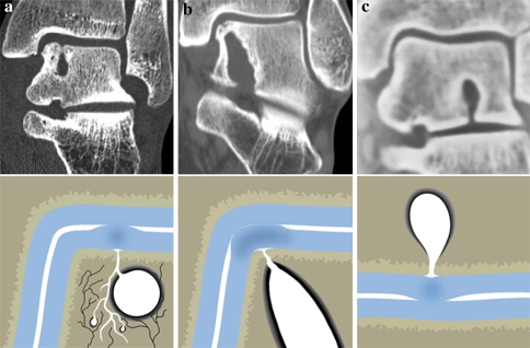
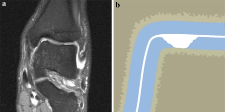
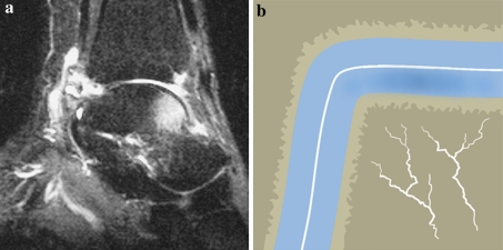
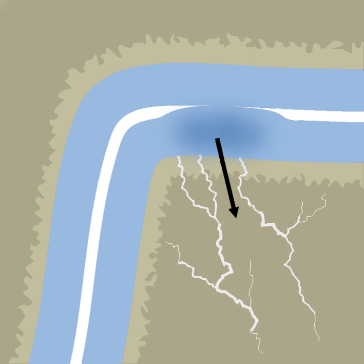
Osteochondral defects of the ankle can either heal and remain asymptomatic or progress to deep ankle pain on weight bearing and formation of subchondral bone cysts. The development of a symptomatic OD depends on various factors, including the damage and insufficient repair of the subchondral bone plate. The ankle joint has a high congruency. During loading, compressed cartilage forces its water into the microfractured subchondral bone, leading to a localized high increased flow and pressure of fluid in the subchondral bone. This will result in local osteolysis and can explain the slow development of a subchondral cyst. The pain does not arise from the cartilage lesion, but is most probably caused by repetitive high fluid pressure during walking, which results in stimulation of the highly innervated subchondral bone underneath the cartilage defect. Understanding the natural history of osteochondral defects could lead to the development of strategies for preventing progressive joint damage.

摘要

踝关节的软骨下骨缺损要么自行愈合且无症状,要么进展为负重时踝关节深部疼痛和形成软骨下骨囊肿。有症状的 OD 的发展取决于多种因素,包括软骨下骨板的损伤和修复不足。踝关节具有高度的一致性。在负重时,受压的软骨将其水分压入微骨折的软骨下骨,导致软骨下骨中的局部高流量和高压流体增加。这将导致局部溶骨,并可以解释软骨下囊肿的缓慢发展。疼痛不是来自软骨病变,而是很可能是由于行走时反复的高流体压力,导致在软骨缺损下方的神经丰富的软骨下骨受到刺激。了解软骨下骨缺损的自然病程可能会导致制定预防关节进行性损伤的策略。

https://cdn.ncbi.nlm.nih.gov/pmc/blobs/4a64/2855020/fe98bc50c141/167_2010_1064_Fig10_HTML.jpg
https://cdn.ncbi.nlm.nih.gov/pmc/blobs/4a64/2855020/ff83bc77ec4c/167_2010_1064_Fig1_HTML.jpg
https://cdn.ncbi.nlm.nih.gov/pmc/blobs/4a64/2855020/2024377f0e0a/167_2010_1064_Fig2_HTML.jpg
https://cdn.ncbi.nlm.nih.gov/pmc/blobs/4a64/2855020/c855416bcae2/167_2010_1064_Fig3_HTML.jpg
https://cdn.ncbi.nlm.nih.gov/pmc/blobs/4a64/2855020/92c76eff68a0/167_2010_1064_Fig4_HTML.jpg
https://cdn.ncbi.nlm.nih.gov/pmc/blobs/4a64/2855020/b8d182cf7f4a/167_2010_1064_Fig5_HTML.jpg
https://cdn.ncbi.nlm.nih.gov/pmc/blobs/4a64/2855020/82b9d51b0603/167_2010_1064_Fig6_HTML.jpg
https://cdn.ncbi.nlm.nih.gov/pmc/blobs/4a64/2855020/6422d36c7d6c/167_2010_1064_Fig7_HTML.jpg
https://cdn.ncbi.nlm.nih.gov/pmc/blobs/4a64/2855020/7be36fd67fea/167_2010_1064_Fig8_HTML.jpg
https://cdn.ncbi.nlm.nih.gov/pmc/blobs/4a64/2855020/9a936bf0c857/167_2010_1064_Fig9_HTML.jpg
https://cdn.ncbi.nlm.nih.gov/pmc/blobs/4a64/2855020/fe98bc50c141/167_2010_1064_Fig10_HTML.jpg

相似文献

1
Osteochondral defects in the ankle: why painful?踝关节的软骨骨缺损:为何会疼痛?
Knee Surg Sports Traumatol Arthrosc. 2010 May;18(5):570-80. doi: 10.1007/s00167-010-1064-x. Epub 2010 Feb 12.
2
The natural history of osteochondral lesions in the ankle.踝关节骨软骨损伤的自然病史。
Instr Course Lect. 2010;59:375-86.
3
Is retrograde drilling really useful for osteochondral lesion of talus with subchondral cyst?: A case report.逆行钻孔术对伴有软骨下囊肿的距骨骨软骨损伤真的有用吗?:一例病例报告。
Medicine (Baltimore). 2016 Dec;95(49):e5418. doi: 10.1097/MD.0000000000005418.
4
Histomorphometric, CT arthrographic, and biomechanical mapping of the human ankle.人体踝关节的组织形态计量学、CT 关节造影和生物力学图谱。
Foot Ankle Int. 2013 Jul;34(7):1025-34. doi: 10.1177/1071100713477636. Epub 2013 Feb 8.
5
Osteochondral lesion of the talus: could age be an indication for arthroscopic treatment?距骨骨软骨损伤:年龄是否是关节镜治疗的指征?
Am J Sports Med. 2012 Feb;40(2):419-24. doi: 10.1177/0363546511423739. Epub 2011 Oct 7.
6
Articular cartilage. Anatomy, injury, and repair.关节软骨。解剖、损伤与修复。
Clin Podiatr Med Surg. 2001 Jan;18(1):35-53.
7
Parameters influencing complaints and joint function in patients with osteochondral lesions of the ankle-an investigation based on data from the German Cartilage Registry (KnorpelRegister DGOU).影响踝关节骨软骨损伤患者主诉及关节功能的因素——基于德国软骨注册中心(KnorpelRegister DGOU)数据的调查
Arch Orthop Trauma Surg. 2017 Mar;137(3):367-373. doi: 10.1007/s00402-017-2638-6. Epub 2017 Feb 6.
8
Outcome of autologous bone grafting with preservation of articular cartilage to treat osteochondral lesions of the talus with large associated subchondral cysts.自体骨移植联合关节软骨保留治疗伴有大的软骨下骨囊肿的距骨骨软骨病变的疗效。
Bone Joint J. 2018 May 1;100-B(5):590-595. doi: 10.1302/0301-620X.100B5.BJJ-2017-1119.R1.
9
Differences in joint morphology between the knee and ankle affect the repair of osteochondral defects in a rabbit model.兔模型中,膝关节与踝关节之间关节形态的差异会影响骨软骨缺损的修复。
J Orthop Surg Res. 2016 Oct 4;11(1):110. doi: 10.1186/s13018-016-0444-4.
10
Management of Subchondral Lesions in the Foot and Ankle.足部和踝关节的软骨下病变的处理。
Clin Podiatr Med Surg. 2023 Jul;40(3):553-568. doi: 10.1016/j.cpm.2023.03.005. Epub 2023 May 3.

引用本文的文献

1
Subchondral bone conditions influence pain in patients with osteochondral lesion of the talus.距骨软骨下骨状况会影响距骨骨软骨损伤患者的疼痛。
Arch Orthop Trauma Surg. 2025 Jun 9;145(1):341. doi: 10.1007/s00402-025-05956-z.
2
Casting and rehabilitation versus skillful neglect for osteochondral lesions of the talus in the pediatric population: the care study, a multicenter, prospective comparative study.小儿距骨骨软骨损伤的石膏固定与康复治疗对比巧妙忽视:CARE研究,一项多中心前瞻性对比研究
BMC Musculoskelet Disord. 2025 May 15;26(1):481. doi: 10.1186/s12891-025-08646-4.
3
The effect of smoking on the outcomes of arthroscopic microfracture for osteochondral lesions of the talus.

本文引用的文献

1
Pressure distribution at the ankle joint.踝关节处的压力分布。
Clin Biomech (Bristol). 1990 Aug;5(3):153-61. doi: 10.1016/0268-0033(90)90018-2.
2
Osteochondritis dissecans of the talus.距骨剥脱性骨软骨炎
J Bone Joint Surg Am. 1947 Jul;29(3):697-706.
3
Treatment of osteochondral lesions of the talus: a systematic review.距骨骨软骨病变的治疗:系统评价。
吸烟对距骨骨软骨损伤关节镜下微骨折治疗效果的影响。
PLoS One. 2025 Apr 22;20(4):e0321894. doi: 10.1371/journal.pone.0321894. eCollection 2025.
4
Surgical technique of an innovative patient-specific metal implant for talar osteochondral lesions.一种用于距骨骨软骨损伤的创新型定制金属植入物的手术技术。
J Exp Orthop. 2025 Apr 7;12(2):e70085. doi: 10.1002/jeo2.70085. eCollection 2025 Apr.
5
Long-Term Results after Autologous Matrix-Induced Chondrogenesis for Osteochondral Lesions of the Talus: A 10-Year Cohort Study.距骨骨软骨损伤自体基质诱导软骨形成术后的长期结果:一项10年队列研究
Cartilage. 2024 Dec 12:19476035241301896. doi: 10.1177/19476035241301896.
6
Higher Accuracy of Arthroscopy Compared to MRI in the Diagnosis of Chondral Lesions in Acute Ankle Fractures: A Prospective Study.关节镜检查在急性踝关节骨折软骨损伤诊断中较磁共振成像具有更高的准确性:一项前瞻性研究
Diagnostics (Basel). 2024 Aug 20;14(16):1810. doi: 10.3390/diagnostics14161810.
7
Effects of Preoperative Abnormality of Posterior Tibial Tendon on the Surgical Outcomes of Medial Osteochondral Lesion of the Talus.术前胫后肌腱异常对距骨内侧骨软骨损伤手术疗效的影响
Cureus. 2024 Jun 10;16(6):e62046. doi: 10.7759/cureus.62046. eCollection 2024 Jun.
8
The MOCART (magnetic resonance observation of cartilage repair tissue) 2.0 Ankle Score.MOCART(软骨修复组织的磁共振观察)2.0踝关节评分
Insights Imaging. 2024 May 31;15(1):126. doi: 10.1186/s13244-024-01696-7.
9
Arthroscopic debridement and microfracture for bilateral osteochondral lesions on the lateral process of the talus involving the subtalar joint: A case report.关节镜下清理术和微骨折术治疗累及跗骨后外侧关节的双侧距骨骨软骨病变:病例报告。
Medicine (Baltimore). 2024 May 24;103(21):e38302. doi: 10.1097/MD.0000000000038302.
10
Improvement in Clinical and Radiographic Outcomes After Isolated Realignment Surgery in Patients With Large Cystic Osteochondral Lesion of the Talar Shoulder and Concurrent Malalignment.距骨肩部大囊性骨软骨损伤并伴有对线不良患者单纯矫正手术后的临床和影像学结果改善
Orthop J Sports Med. 2024 Apr 10;12(4):23259671241237126. doi: 10.1177/23259671241237126. eCollection 2024 Apr.
Knee Surg Sports Traumatol Arthrosc. 2010 Feb;18(2):238-46. doi: 10.1007/s00167-009-0942-6. Epub 2009 Oct 27.
4
Histologic findings and possible causes of osteochondritis dissecans of the knee.膝关节剥脱性骨软骨炎的组织学发现及可能病因。
Am J Sports Med. 2009 Oct;37(10):2003-8. doi: 10.1177/0363546509346542. Epub 2009 Sep 8.
5
Bilateral osteochondral lesions of the talus.距骨双侧骨软骨损伤
Foot Ankle Int. 2009 Aug;30(8):723-7. doi: 10.3113/FAI.2009.0723.
6
Bone resorption induced by fluid flow.流体流动诱导的骨吸收。
J Biomech Eng. 2009 Sep;131(9):094505. doi: 10.1115/1.3194756.
7
Arthroscopic treatment of chronic osteochondral lesions of the talus: long-term results.关节镜治疗距骨慢性骨软骨损伤:长期疗效
Am J Sports Med. 2008 Sep;36(9):1750-62. doi: 10.1177/0363546508316773.
8
In vivo cartilage contact deformation of human ankle joints under full body weight.人体踝关节在全身重量下的体内软骨接触变形。
J Orthop Res. 2008 Aug;26(8):1081-9. doi: 10.1002/jor.20593.
9
A stereophotographic study of ankle joint contact area.踝关节接触面积的立体摄影研究。
J Orthop Res. 2007 Nov;25(11):1465-73. doi: 10.1002/jor.20425.
10
Osteochondral lesions of the talus: localization and morphologic data from 424 patients using a novel anatomical grid scheme.距骨骨软骨损伤:采用新型解剖网格方案对424例患者的损伤部位及形态学数据进行分析
Foot Ankle Int. 2007 Feb;28(2):154-61. doi: 10.3113/FAI.2007.0154.