Darwich Ali, Nörenberg Dominik, Adam Julia, Hetjens Svetlana, Bdeir Mohamad, Schilder Andreas, Thier Steffen, Gravius Sascha, Jawhar Ahmed
Department of Orthopedic and Trauma Surgery, University Medical Centre Mannheim, Medical Faculty Mannheim, University of Heidelberg, Theodor-Kutzer-Ufer 1-3, 68167 Mannheim, Germany.
Department of Radiology and Nuclear Medicine, University Medical Centre Mannheim, Medical Faculty Mannheim, University of Heidelberg, Theodor-Kutzer-Ufer 1-3, 68167 Mannheim, Germany.
Diagnostics (Basel). 2024 Aug 20;14(16):1810. doi: 10.3390/diagnostics14161810.
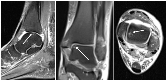
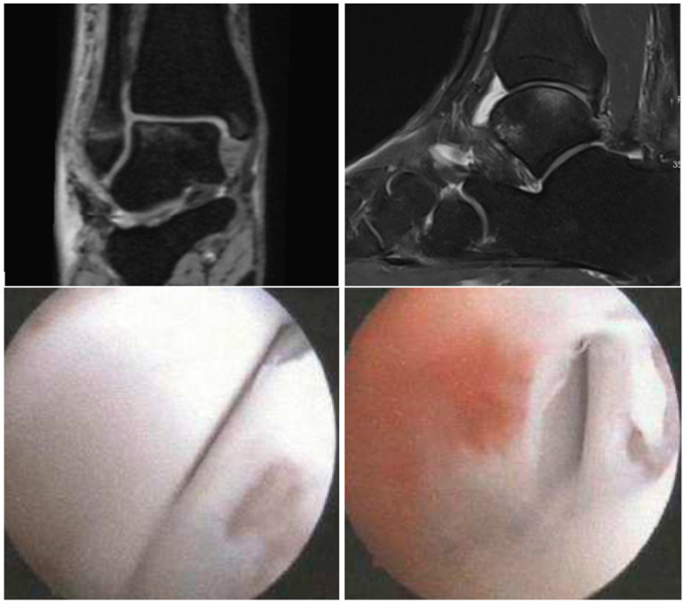
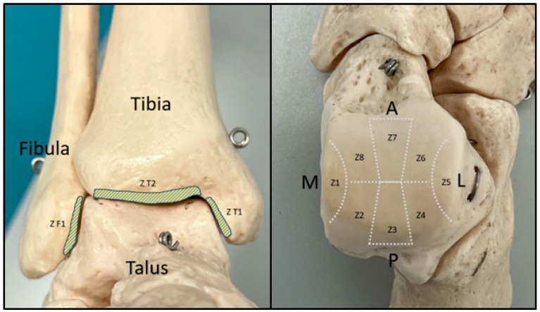
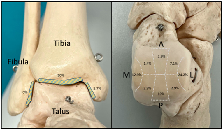
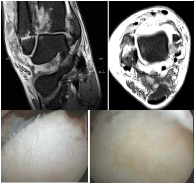
Even after successful surgery for acute ankle fractures, many patients continue having complaints. A possible explanation is the presence of concomitant chondral lesions. The aim of this study is to investigate the accuracy of MRI compared to that of arthroscopy in the assessment of chondral lesions in acute ankle fractures. In this prospective single-center study, patients presenting with acute ankle fractures over a period of three years were identified. A preoperative MRI was performed within a maximum of 10 days after trauma. During surgery, ankle arthroscopy was also performed. The International Cartilage Repair Society (ICRS) cartilage lesion classification was used to grade the detected chondral lesions. To localize the chondral lesions, the talar dome was divided into eight zones and the tibial/fibular articular surfaces into three zones. In total, 65 patients (28 females) with a mean age of 41.1 ± 15 years were included. In the MRI scans, 70 chondral lesions were detected (69.2% of patients) affecting mostly the tibial plafond (30%) and mostly graded as ICRS 3. The mean lesion area measured was 20.8 mm. In the arthroscopy, 85 chondral lesions were detected (70.8% of patients) affecting mostly the medial surface of the talar dome (25.9%) and mostly graded ICRS 3. The mean lesion area measured was 43.4 mm. The highest agreement between the two methods was observed in the size estimation of the chondral lesions. The present study shows the reduced accuracy of MRI when compared to arthroscopy in the assessment of traumatic chondral lesions in the setting of acute ankle fractures especially regarding lesion size. MRI remains an essential instrument in the evaluation of such lesions; however, surgeons should take this discrepancy into consideration, particularly the underestimation of chondral lesions' size in the preoperative planning of surgical treatment and operative technique.
即使急性踝关节骨折手术成功,许多患者仍有不适。一个可能的解释是存在合并软骨损伤。本研究的目的是调查在评估急性踝关节骨折软骨损伤时,磁共振成像(MRI)与关节镜检查相比的准确性。在这项前瞻性单中心研究中,确定了三年内出现急性踝关节骨折的患者。在创伤后最多10天内进行术前MRI检查。手术期间,也进行了踝关节镜检查。采用国际软骨修复协会(ICRS)软骨损伤分类对检测到的软骨损伤进行分级。为了定位软骨损伤,将距骨穹顶分为八个区域,胫骨/腓骨关节面分为三个区域。总共纳入了65例患者(28例女性),平均年龄为41.1±15岁。在MRI扫描中,检测到70处软骨损伤(占患者的69.2%),主要影响胫骨平台(30%),大多分级为ICRS 3级。测得的平均损伤面积为20.8平方毫米。在关节镜检查中,检测到85处软骨损伤(占患者的70.8%),主要影响距骨穹顶的内表面(25.9%),大多分级为ICRS 3级。测得的平均损伤面积为43.4平方毫米。两种方法之间在软骨损伤大小估计方面的一致性最高。本研究表明,在评估急性踝关节骨折情况下的创伤性软骨损伤时,尤其是在损伤大小方面,MRI与关节镜检查相比准确性较低。MRI仍然是评估此类损伤的重要工具;然而,外科医生应考虑到这种差异,特别是在手术治疗和手术技术的术前规划中软骨损伤大小被低估的情况。