NSE MRI Unit, National Society for Epilepsy, Chalfont St Peter, UK.
Brain. 2010 Apr;133(Pt 4):1186-99. doi: 10.1093/brain/awq006. Epub 2010 Feb 15.
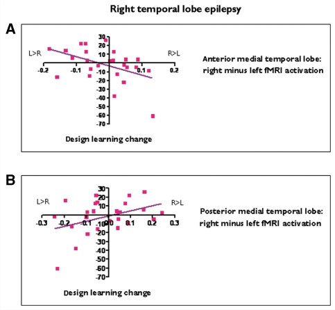
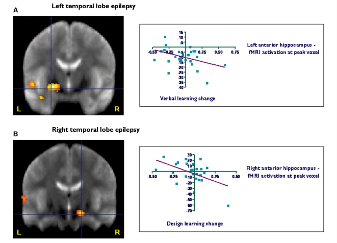
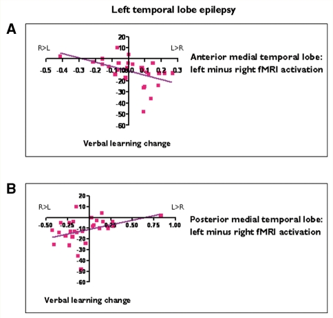
Functional magnetic resonance imaging can demonstrate the functional anatomy of cognitive processes. In patients with refractory temporal lobe epilepsy, evaluation of preoperative verbal and visual memory function is important as anterior temporal lobe resections may result in material specific memory impairment, typically verbal memory decline following left and visual memory decline after right anterior temporal lobe resection. This study aimed to investigate reorganization of memory functions in temporal lobe epilepsy and to determine whether preoperative memory functional magnetic resonance imaging may predict memory changes following anterior temporal lobe resection. We studied 72 patients with unilateral medial temporal lobe epilepsy (41 left) and 20 healthy controls. A functional magnetic resonance imaging memory encoding paradigm for pictures, words and faces was used testing verbal and visual memory in a single scanning session on a 3T magnetic resonance imaging scanner. Fifty-four patients subsequently underwent left (29) or right (25) anterior temporal lobe resection. Verbal and design learning were assessed before and 4 months after surgery. Event-related functional magnetic resonance imaging analysis revealed that in left temporal lobe epilepsy, greater left hippocampal activation for word encoding correlated with better verbal memory. In right temporal lobe epilepsy, greater right hippocampal activation for face encoding correlated with better visual memory. In left temporal lobe epilepsy, greater left than right anterior hippocampal activation on word encoding correlated with greater verbal memory decline after left anterior temporal lobe resection, while greater left than right posterior hippocampal activation correlated with better postoperative verbal memory outcome. In right temporal lobe epilepsy, greater right than left anterior hippocampal functional magnetic resonance imaging activation on face encoding predicted greater visual memory decline after right anterior temporal lobe resection, while greater right than left posterior hippocampal activation correlated with better visual memory outcome. Stepwise linear regression identified asymmetry of activation for encoding words and faces in the ipsilateral anterior medial temporal lobe as strongest predictors for postoperative verbal and visual memory decline. Activation asymmetry, language lateralization and performance on preoperative neuropsychological tests predicted clinically significant verbal memory decline in all patients who underwent left anterior temporal lobe resection, but were less able to predict visual memory decline after right anterior temporal lobe resection. Preoperative memory functional magnetic resonance imaging was the strongest predictor of verbal and visual memory decline following anterior temporal lobe resection. Preoperatively, verbal and visual memory function utilized the damaged, ipsilateral hippocampus and also the contralateral hippocampus. Memory function in the ipsilateral posterior hippocampus may contribute to better preservation of memory after surgery.
功能磁共振成像可以显示认知过程的功能解剖结构。在难治性颞叶癫痫患者中,术前评估言语和视觉记忆功能非常重要,因为前颞叶切除术可能导致特定材料记忆障碍,通常左前颞叶切除后出现言语记忆下降,右前颞叶切除后出现视觉记忆下降。本研究旨在探讨颞叶癫痫患者记忆功能的重组,并确定术前记忆功能磁共振成像是否可以预测前颞叶切除术后的记忆变化。我们研究了 72 例单侧内侧颞叶癫痫患者(41 例左侧)和 20 名健康对照者。使用功能磁共振成像记忆编码范式对图片、单词和面孔进行测试,在 3T 磁共振成像扫描仪上单次扫描中测试言语和视觉记忆。54 例患者随后接受了左(29 例)或右(25 例)前颞叶切除术。手术前和手术后 4 个月评估言语和设计学习。事件相关功能磁共振成像分析显示,在左侧颞叶癫痫中,单词编码时左侧海马体的激活程度与言语记忆的改善相关。在右侧颞叶癫痫中,面孔编码时右侧海马体的激活程度与视觉记忆的改善相关。在左侧颞叶癫痫中,单词编码时左侧前海马体的激活程度大于右侧与左前颞叶切除术后言语记忆下降程度较大相关,而左侧后海马体的激活程度大于右侧与术后言语记忆结局较好相关。在右侧颞叶癫痫中,面孔编码时右侧前海马体的激活程度大于左侧与右前颞叶切除术后视觉记忆下降程度较大相关,而右侧后海马体的激活程度大于左侧与视觉记忆结局较好相关。逐步线性回归确定了同侧前内侧颞叶中单词和面孔编码的激活不对称性是术后言语和视觉记忆下降的最强预测因子。激活不对称性、语言偏侧性和术前神经心理学测试的表现预测了所有接受左前颞叶切除术的患者的临床显著言语记忆下降,但对右前颞叶切除术后的视觉记忆下降预测能力较弱。术前记忆功能磁共振成像是前颞叶切除术后言语和视觉记忆下降的最强预测因子。术前,言语和视觉记忆功能既利用了受损的同侧海马体,也利用了对侧海马体。同侧后海马体的记忆功能可能有助于手术后记忆的更好保留。