Department of Pathology, Biological Sciences Institute, Federal University of Minas Gerais, Av. Antônio Carlos, 6627, Belo Horizonte, Minas Gerais, 31270-901, Brazil.
BMC Cancer. 2010 Feb 23;10:61. doi: 10.1186/1471-2407-10-61.
It has been suggested that columnar cell lesions indicate an alteration of the human mammary gland involved in the development of breast cancer. They have not previously been described in canine mammary gland. The aim of this paper is describe the morphologic spectrum of columnar cell lesions in canine mammary gland specimens and their association with other breast lesions.
A total of 126 lesions were subjected to a comprehensive morphological review based upon the human breast classification system for columnar cell lesions. The presence of preinvasive (epithelial hyperplasia and in situ carcinoma) and invasive lesions was determined and immunophenotypic analysis (estrogen receptor (ER), progesterone receptor (PgR), high molecular weight cytokeratin (34betaE-12), E-cadherin, Ki-67, HER-2 and P53) was perfomed.
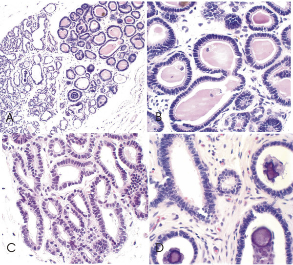
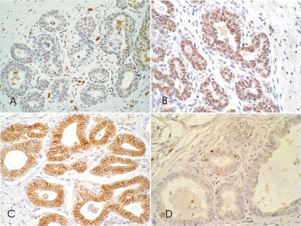
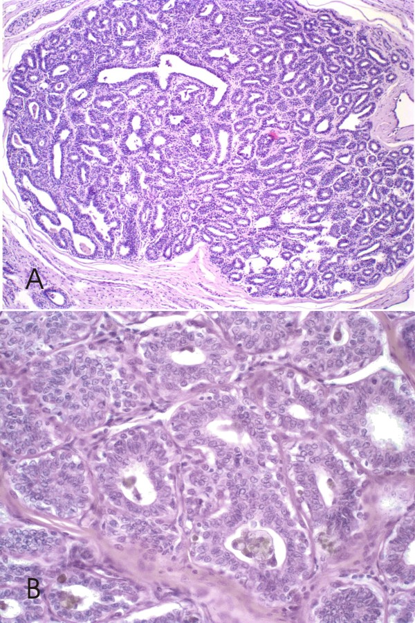
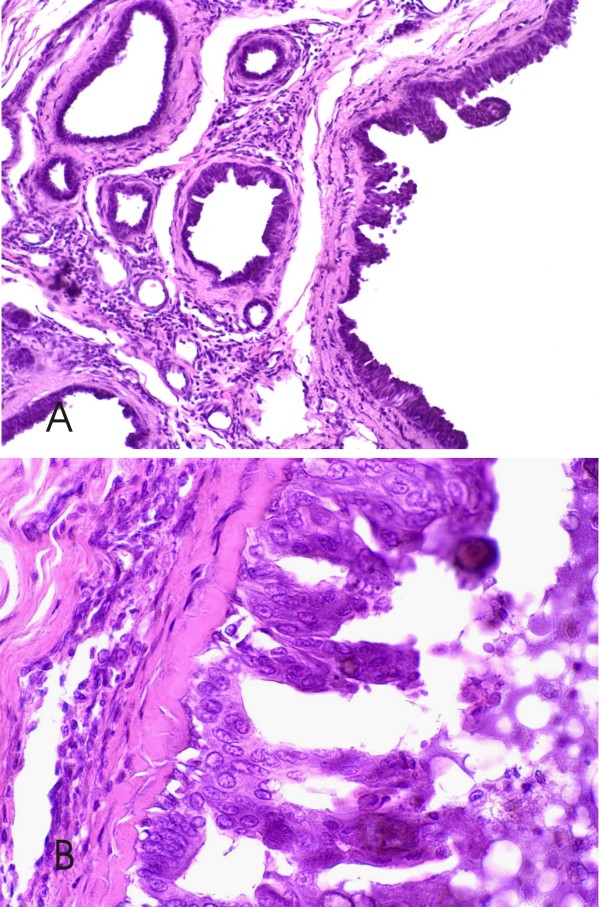
Columnar cell lesions were identified in 67 (53.1%) of the 126 canine mammary glands with intraepithelial alterations. They were observed in the terminal duct lobular units and characterized at dilated acini may be lined by several layers of columnar epithelial cells with elongated nuclei. Of the columnar cell lesions identified, 41 (61.2%) were without and 26 (38.8%) with atypia. Association with ductal hyperplasia was observed in 45/67 (67.1%). Sixty (89.5%) of the columnar cell lesions coexisted with neoplastic lesions (20 in situ carcinomas, 19 invasive carcinomas and 21 benign tumors). The columnar cells were ER, PgR and E-cadherin positive but negative for cytokeratin 34betaE-12, HER-2 and P53. The proliferation rate as measured by Ki-67 appeared higher in the lesions analyzed than in normal TDLUs.
Columnar cell lesions in canine mammary gland are pathologically and immunophenotypically similar to those in human breast. This may suggest that dogs are a suitable model for the comparative study of noninvasive breast lesions.
有研究表明柱状细胞病变表明乳腺发生了与乳腺癌发展相关的改变。此前,犬乳腺中并未描述过此类病变。本文旨在描述犬乳腺柱状细胞病变的形态学谱及其与其他乳腺病变的关系。
对 126 例病变进行了全面的形态学回顾,基于人乳腺柱状细胞病变分类系统进行分类。确定了是否存在浸润前病变(上皮增生和原位癌)和浸润性病变,并进行了免疫表型分析(雌激素受体(ER)、孕激素受体(PgR)、高分子量细胞角蛋白(34βE-12)、E-钙黏蛋白、Ki-67、HER-2 和 P53)。
在 126 例犬乳腺中有 67 例(53.1%)存在上皮内改变的柱状细胞病变。这些病变位于终末导管小叶单位,表现为扩张的腺泡可能由数层柱状上皮细胞组成,细胞核拉长。在鉴定的柱状细胞病变中,无异型性的有 41 例(61.2%),有异型性的有 26 例(38.8%)。与导管增生相关的有 45/67(67.1%)例。60 例(89.5%)柱状细胞病变与肿瘤病变共存(20 例原位癌、19 例浸润性癌和 21 例良性肿瘤)。柱状细胞 ER、PgR 和 E-钙黏蛋白阳性,而细胞角蛋白 34βE-12、HER-2 和 P53 阴性。Ki-67 测量的增殖率在分析的病变中高于正常 TDLU。
犬乳腺柱状细胞病变在病理和免疫表型上与人乳腺相似。这表明犬是研究非浸润性乳腺病变的合适模型。