Kan Akinori, Oshida Midori, Oshida Shigemi, Imada Masato, Nakagawa Takumi, Okinaga Shuji
Department of Sensory & Motor System Medicine, Faculty of Medicine, University of Tokyo, 7-3-1 Hongo, Bunkyo, Tokyo 113-8655, Japan.
Sports Med Arthrosc Rehabil Ther Technol. 2010 Jan 12;2:1. doi: 10.1186/1758-2555-2-1.
Traumatic injury and surgical meniscectomy of a medial meniscus are known to cause subsequent knee osteoarthritis. However, the difference in the prevalence of osteoarthritis caused by the individual type of the medial meniscal tear has not been elucidated. The aim of this study was to investigate what type of tear is predominantly responsible for the degradation of articular cartilage in the medial compartment of knee joints.
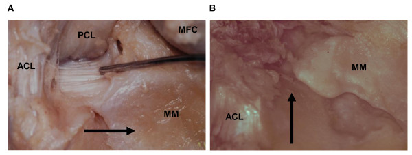
Five hundred and forty eight cadaveric knees (290 male and 258 female) were registered in this study. The average age of cadavers at death was 78.8 years old (range: 52-103 years). The knees were macroscopically examined and their medial menisci were classified into four groups according to types of tears: "no tear", "radial tear of posterior horn", "other types of tear" and "worn-out meniscus" groups. The severity of cartilage degradation in their medial compartment of knee joints was evaluated using the international cartilage repair society (ICRS) grading system. We statistically compared the ICRS grades among the groups using Mann-Whitney U test.
The knees were assigned into the four groups: 416 "no tear" knees, 51 "radial tear of posterior horn" knees, 71 "other types of tear" knees, and 10 "worn-out meniscus" knees. The knees with substantial meniscal tears showed the severer ICRS grades of cartilage degradation than those without meniscal tears. In addition, the ICRS grades were significantly severer in the "radial tear of posterior horn" group than in the "other types of tear" group, suggesting that the radial tear of posterior horn in the medial meniscus is one of the risk factors for cartilage degradation of joint surface.
We have clarified the relationship between the radial tear of posterior horn in the medial meniscus and the severer grade of cartilage degradation. This study indicates that the efforts should be made to restore the anatomical role of the posterior horn in keeping the hoop strain, when patients' physical activity levels are high and the tear pattern is simple enough to be securely sutured.
创伤性损伤和内侧半月板手术切除已知会导致随后的膝关节骨关节炎。然而,内侧半月板撕裂的个体类型所导致的骨关节炎患病率差异尚未阐明。本研究的目的是调查哪种类型的撕裂是膝关节内侧间室关节软骨退变的主要原因。
本研究纳入了548具尸体膝关节(男性290例,女性258例)。尸体死亡时的平均年龄为78.8岁(范围:52 - 103岁)。对膝关节进行宏观检查,并根据撕裂类型将其内侧半月板分为四组:“无撕裂”、“后角放射状撕裂”、“其他类型撕裂”和“磨损半月板”组。使用国际软骨修复协会(ICRS)分级系统评估膝关节内侧间室软骨退变的严重程度。我们使用Mann-Whitney U检验对各组间的ICRS分级进行统计学比较。
膝关节被分为四组:416例“无撕裂”膝关节、51例“后角放射状撕裂”膝关节、71例“其他类型撕裂”膝关节和10例“磨损半月板”膝关节。有明显半月板撕裂的膝关节显示出比无半月板撕裂的膝关节更严重的ICRS软骨退变分级。此外,“后角放射状撕裂”组的ICRS分级明显比“其他类型撕裂”组更严重,表明内侧半月板后角放射状撕裂是关节面软骨退变的危险因素之一。
我们已经阐明了内侧半月板后角放射状撕裂与更严重的软骨退变分级之间的关系。本研究表明,当患者身体活动水平较高且撕裂模式简单到足以安全缝合时,应努力恢复后角在维持环向应变方面的解剖学作用。