• 文献检索
  • 文档翻译
  • 深度研究
  • 学术资讯
  • Suppr Zotero 插件Zotero 插件
  • 邀请有礼
  • 套餐&价格
  • 历史记录
应用&插件
Suppr Zotero 插件Zotero 插件浏览器插件Mac 客户端Windows 客户端微信小程序
定价
高级版会员购买积分包购买API积分包
服务
文献检索文档翻译深度研究API 文档MCP 服务
关于我们
关于 Suppr公司介绍联系我们用户协议隐私条款
关注我们

Suppr 超能文献

核心技术专利:CN118964589B侵权必究
粤ICP备2023148730 号-1Suppr @ 2026

文献检索

告别复杂PubMed语法,用中文像聊天一样搜索,搜遍4000万医学文献。AI智能推荐,让科研检索更轻松。

立即免费搜索

文件翻译

保留排版,准确专业,支持PDF/Word/PPT等文件格式,支持 12+语言互译。

免费翻译文档

深度研究

AI帮你快速写综述,25分钟生成高质量综述,智能提取关键信息,辅助科研写作。

立即免费体验

一种诊断内侧半月板后根撕裂的新临床体征。

A New Clinical Sign for Diagnosing Medial Meniscus Posterior Root Tear.

作者信息

Akmese Ramazan, Malatyalı Batu, Kocaoglu Hakan, Akkaya Zehra, Kalem Mahmut

机构信息

Department of Orthopedics and Traumatology, Ankara University Faculty of Medicine, Ankara, Turkey.

Department of Radiology, Ankara University Faculty of Medicine, Ankara, Turkey.

出版信息

Orthop J Sports Med. 2021 Jan 22;9(1):2325967120975511. doi: 10.1177/2325967120975511. eCollection 2021 Jan.

DOI:10.1177/2325967120975511
PMID:33553453
原文链接:https://pmc.ncbi.nlm.nih.gov/articles/PMC7841682/
Abstract

BACKGROUND

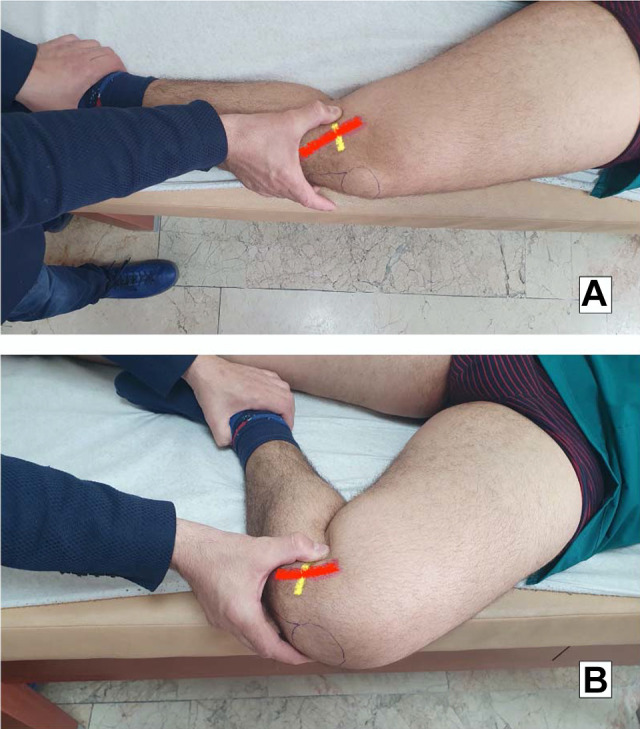
In the presence of medial meniscus posterior root tear (MMPRT), there is a possibility of reduced compression of meniscal tissue in hyperflexion as the intra-articular mobility of the meniscus increases. This phenomenon can be mimicked during clinical examination.

PURPOSE

To describe, evaluate, and validate the diagnostic performance of a new clinical indicator, the Akmese sign, for the diagnosis of an MMPRT.

STUDY DESIGN

Cohort study (diagnosis); Level of evidence, 2.

METHODS

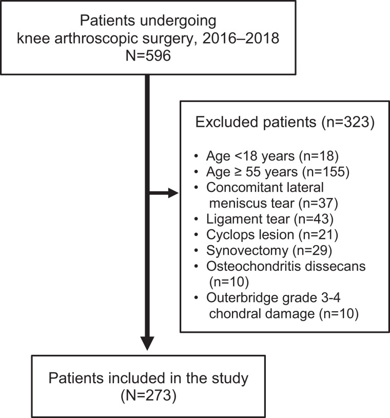
In this study, we prospectively enrolled patients aged 18 to 55 years who were scheduled for arthroscopic surgery after a diagnosis of medial meniscal lesion at a single institution between January 2016 and January 2018. All of the patients underwent preoperative examination for the Akmese sign. All surgeries were performed by a single surgeon with more than 5 years of experience in sports injury surgery, who was blinded to the Akmese sign results.

RESULTS

A total of 273 patients with a mean age of 42.4 ± 5.3 years met the study criteria. The Akmese sign was identified as positive in 33 patients, and MMPRT was confirmed during arthroscopy in 36 cases. The performance parameters of the Akmese sign were a sensitivity of 86.1%, specificity of 99.1%, Youden index of 0.85, and kappa index of 0.88.

CONCLUSION

This study showed that the Akmese sign is a useful new physical examination test that can help clinicians distinguish MMPRTs from other meniscal medial meniscal pathology.

摘要

背景

在内侧半月板后根部撕裂(MMPRT)的情况下,随着半月板在关节内的活动度增加,在极度屈曲时半月板组织的压缩可能会降低。这种现象在临床检查中可能会被模拟出来。

目的

描述、评估和验证一种新的临床指标——阿克梅斯征(Akmese sign)对MMPRT的诊断性能。

研究设计

队列研究(诊断);证据等级,2级。

方法

在本研究中,我们前瞻性纳入了2016年1月至2018年1月期间在单一机构被诊断为内侧半月板损伤后计划接受关节镜手术的18至55岁患者。所有患者均接受了阿克梅斯征的术前检查。所有手术均由一位在运动损伤手术方面有超过5年经验的外科医生进行,该医生对阿克梅斯征的结果不知情。

结果

共有273例平均年龄为42.4±5.3岁的患者符合研究标准。33例患者的阿克梅斯征被判定为阳性,36例在关节镜检查中确诊为MMPRT。阿克梅斯征的性能参数为敏感性86.1%、特异性99.1%、约登指数0.85和kappa指数0.88。

结论

本研究表明,阿克梅斯征是一种有用的新体格检查方法,可帮助临床医生将MMPRT与其他内侧半月板病变区分开来。

https://cdn.ncbi.nlm.nih.gov/pmc/blobs/4a53/7841682/9ec94d4ef446/10.1177_2325967120975511-fig2.jpg
https://cdn.ncbi.nlm.nih.gov/pmc/blobs/4a53/7841682/9d0034bd78e0/10.1177_2325967120975511-fig1.jpg
https://cdn.ncbi.nlm.nih.gov/pmc/blobs/4a53/7841682/9ec94d4ef446/10.1177_2325967120975511-fig2.jpg
https://cdn.ncbi.nlm.nih.gov/pmc/blobs/4a53/7841682/9d0034bd78e0/10.1177_2325967120975511-fig1.jpg
https://cdn.ncbi.nlm.nih.gov/pmc/blobs/4a53/7841682/9ec94d4ef446/10.1177_2325967120975511-fig2.jpg

相似文献

1
A New Clinical Sign for Diagnosing Medial Meniscus Posterior Root Tear.一种诊断内侧半月板后根撕裂的新临床体征。
Orthop J Sports Med. 2021 Jan 22;9(1):2325967120975511. doi: 10.1177/2325967120975511. eCollection 2021 Jan.
2
Arthroscopic scoring system of meniscal healing following medial meniscus posterior root repair.关节镜下内侧半月板后根修复后半月板愈合评分系统。
Int Orthop. 2019 May;43(5):1239-1245. doi: 10.1007/s00264-018-4071-z. Epub 2018 Aug 1.
3
[Arthroscopic Finding of Knee Joint in Relation to Age and Its Comparison with Pre-Operative Clinical Finding - a Retrospective Study].[膝关节镜检查结果与年龄的关系及其与术前临床检查结果的比较——一项回顾性研究]
Acta Chir Orthop Traumatol Cech. 2017;84(3):175-181.
4
A giraffe neck sign of the medial meniscus: A characteristic finding of the medial meniscus posterior root tear on magnetic resonance imaging.内侧半月板的长颈鹿颈征:磁共振成像上内侧半月板后根部撕裂的特征性表现。
J Orthop Sci. 2017 Jul;22(4):731-736. doi: 10.1016/j.jos.2017.03.013. Epub 2017 Apr 13.
5
Non-anatomic repair of medial meniscus posterior root tears to the posterior capsule provided favourable outcomes in middle-aged and older patients.对中老年患者后囊内半月板后根撕裂行非解剖修复可获得良好的效果。
Knee Surg Sports Traumatol Arthrosc. 2021 Dec;29(12):4261-4269. doi: 10.1007/s00167-021-06532-9. Epub 2021 Apr 9.
6
The characteristic arthroscopic 'eyelid- turnup' sign indicated for partial-thickness longitudinal medial meniscus tear.特征性的关节镜“眼睑翻转”征提示内侧半月板纵行部分厚度撕裂。
Technol Health Care. 2019;27(6):661-668. doi: 10.3233/THC-191640.
7
An MRI-based suspension bridge sign can predict an arthroscopically favorable meniscal healing following the medial meniscus posterior root repair.基于 MRI 的悬索桥征可预测内侧半月板后根修复术后关节镜下有利的半月板愈合。
J Orthop Sci. 2021 Mar;26(2):237-242. doi: 10.1016/j.jos.2020.03.012. Epub 2020 Apr 10.
8
Early Surgical Repair of Medial Meniscus Posterior Root Tear Minimizes the Progression of Meniscal Extrusion: 2-Year Follow-up of Clinical and Radiographic Parameters After Arthroscopic Transtibial Pull-out Repair.关节镜下经胫骨隧道拉拔修复术后 2 年的临床和影像学参数随访:内侧半月板后根撕裂的早期手术修复可最大限度减少半月板挤出的进展
Am J Sports Med. 2020 Sep;48(11):2692-2702. doi: 10.1177/0363546520940715. Epub 2020 Jul 30.
9
A characteristic MRI finding to diagnose a partial tear of the medial meniscus posterior root: an ocarina sign.诊断内侧半月板后根部部分撕裂的一个典型MRI表现:陶笛征。
Knee Surg Relat Res. 2021 Oct 9;33(1):38. doi: 10.1186/s43019-021-00120-4.
10
[Diagnostic value of MRI for posterior root tear of medial and lateral meniscus].[MRI对内外侧半月板后根撕裂的诊断价值]
Zhongguo Gu Shang. 2018 Mar 25;31(3):263-266. doi: 10.3969/j.issn.1003-0034.2018.03.014.

引用本文的文献

1
Diagnosis and Treatment Strategies of Meniscus Root Tears: A Scoping Review.半月板根部撕裂的诊断与治疗策略:一项范围综述
Orthop J Sports Med. 2024 Nov 1;12(11):23259671241283962. doi: 10.1177/23259671241283962. eCollection 2024 Nov.
2
Medial Meniscus Posterior Root Tear: How Far Have We Come and What Remains?内侧半月板后根撕裂:我们已经走了多远,还有什么要做的?
Medicina (Kaunas). 2023 Jun 21;59(7):1181. doi: 10.3390/medicina59071181.

本文引用的文献

1
STARD 2015 guidelines for reporting diagnostic accuracy studies: explanation and elaboration.《STARD 2015诊断准确性研究报告指南:解释与详述》
BMJ Open. 2016 Nov 14;6(11):e012799. doi: 10.1136/bmjopen-2016-012799.
2
Effect of lateral meniscal root tear on the stability of the anterior cruciate ligament-deficient knee.外侧半月板根部撕裂对前交叉韧带损伤膝关节稳定性的影响。
Am J Sports Med. 2015 Apr;43(4):905-11. doi: 10.1177/0363546514563910. Epub 2015 Jan 14.
3
Meniscal root tears: significance, diagnosis, and treatment.半月板根部撕裂:意义、诊断与治疗
Am J Sports Med. 2014 Dec;42(12):3016-30. doi: 10.1177/0363546514524162. Epub 2014 Mar 12.
4
Posterior root tears of the lateral meniscus.外侧半月板后根部撕裂
Knee Surg Sports Traumatol Arthrosc. 2015 Jan;23(1):119-25. doi: 10.1007/s00167-014-2904-x. Epub 2014 Feb 15.
5
Medial posterior meniscal root tears are associated with development or worsening of medial tibiofemoral cartilage damage: the multicenter osteoarthritis study.内侧后半月板根撕裂与内侧胫股软骨损伤的发展或恶化有关:多中心骨关节炎研究。
Radiology. 2013 Sep;268(3):814-21. doi: 10.1148/radiol.13122544. Epub 2013 May 21.
6
Meniscal root tears: from basic science to ultimate surgery.半月板根部撕裂:从基础科学到终极手术。
Br Med Bull. 2013;106:91-115. doi: 10.1093/bmb/ldt002. Epub 2013 Jan 31.
7
Risk factors for medial meniscus posterior root tear.内侧半月板后根撕裂的危险因素。
Am J Sports Med. 2012 Jul;40(7):1606-10. doi: 10.1177/0363546512447792. Epub 2012 May 11.
8
Meniscus root repair.半月板根部修复
Sports Med Arthrosc Rev. 2012 Jun;20(2):86-94. doi: 10.1097/JSA.0b013e31825186ca.
9
A clinical sign to detect root avulsions of the posterior horn of the medial meniscus.一个用于检测内侧半月板后角根部撕脱的临床征象。
Knee Surg Sports Traumatol Arthrosc. 2011 Dec;19(12):2072-5. doi: 10.1007/s00167-011-1550-9. Epub 2011 May 24.
10
Medial meniscus root tear refixation: comparison of clinical, radiologic, and arthroscopic findings with medial meniscectomy.内侧半月板后根部撕裂修复固定:与内侧半月板切除术的临床、放射学和关节镜检查结果比较。
Arthroscopy. 2011 Mar;27(3):346-54. doi: 10.1016/j.arthro.2010.08.005. Epub 2010 Oct 29.