Dilsiz Alparslan, Aydin Tugba, Gursan Nesrin
Department of Periodontology, Faculty of Dentistry, Ataturk University, 25240, Erzurum/Turkey.
Cases J. 2009 Sep 9;2:8622. doi: 10.1186/1757-1626-0002-0000008622.
Hemangioma is a relatively common benign proliferation of blood vessels that primarily develops during childhood. Two main forms of hemangioma recognized: capillary and cavernous. The capillary form presents as a flat area consisting of numerous small capillaries. Cavernous hemangioma appears as an elevated lesion of a deep red color, and consists of large dilated sinuses filled with blood. The purpose of the study was to report the case of a capillary hemangioma in a patient and to describe the successful treatment of this case.
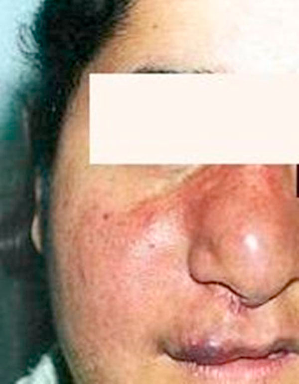
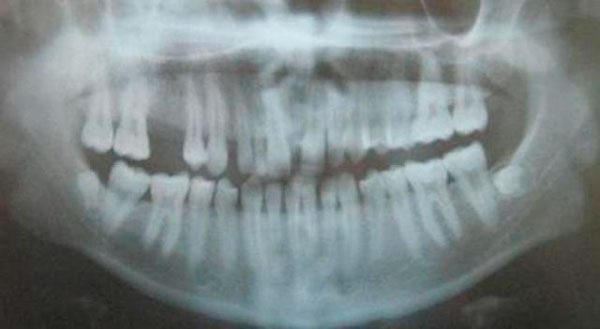
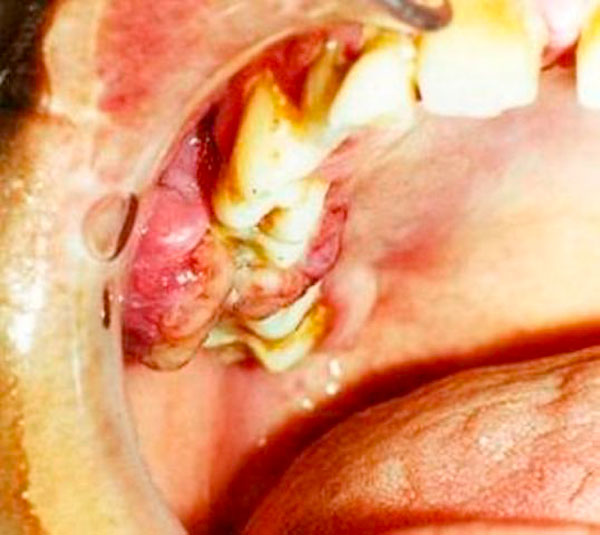
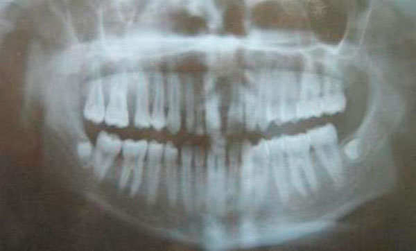
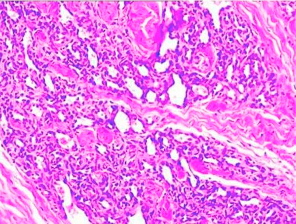
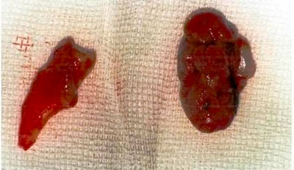
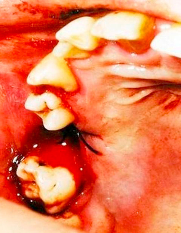
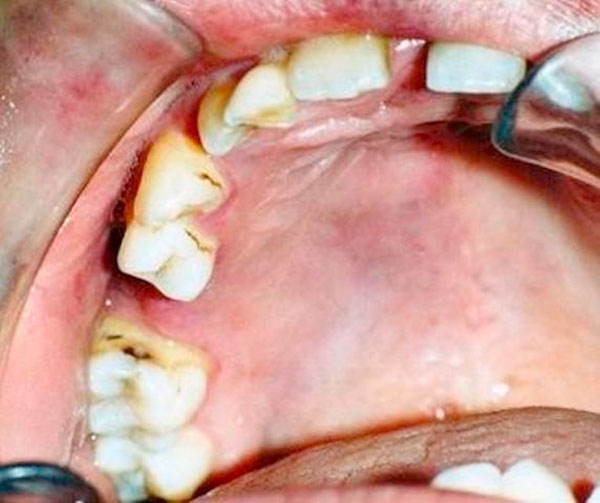
The patient was a 19-year-old female who presented herself to the Atatürk University, Faculty of Dentistry, Department of Periodontology, with the complaint of bleeding and slowly enlarging mass on the upper right molar region. The lesion was diagnosed as capillary hemangioma after clinical examination and biopsy. Treatment consisted of scaling, root planning and surgical excision. Four months after surgery healing was occurred and two years later area of the lesion appeared completely normal as clinically.
The surface is highly keratinized and no further growth was evidenced during the two year of follow-up. Early detection and biopsy is necessary to determine the clinical behavior of the tumor and potential dentoalveolar complications.
血管瘤是一种相对常见的血管良性增生,主要在儿童期发生。血管瘤主要有两种类型:毛细血管型和海绵状型。毛细血管型表现为一个由众多小毛细血管组成的扁平区域。海绵状血管瘤表现为一个深红色的隆起病变,由充满血液的大扩张窦组成。本研究的目的是报告一例患者的毛细血管型血管瘤病例,并描述该病例的成功治疗过程。
患者为一名19岁女性,因右上磨牙区出血及肿物缓慢增大就诊于阿塔图尔克大学牙科学院牙周病科。经临床检查和活检,该病变被诊断为毛细血管型血管瘤。治疗包括洁治、根面平整和手术切除。术后四个月伤口愈合,两年后病变区域临床外观完全正常。
病变表面高度角化,在两年的随访期间未发现进一步生长。早期检测和活检对于确定肿瘤的临床行为和潜在的牙槽并发症是必要的。