Institut für Molekularbiologie, OE5250, Medizinische Hochschule Hannover, Carl-Neuberg-Str.1, D-30625 Hannover, Germany.
Circ Res. 2010 Apr 16;106(7):1212-20. doi: 10.1161/CIRCRESAHA.110.217455. Epub 2010 Feb 25.
The cardiac venous pole is a common focus of congenital malformations and atrial arrhythmias, yet little is known about the cellular and molecular mechanisms that regulate its development. The systemic venous return myocardium (sinus node and sinus horns) forms only late in cardiogenesis from a pool of pericardial mesenchymal precursor cells.
To analyze the cellular and molecular mechanisms directing the formation of the fetal sinus horns.
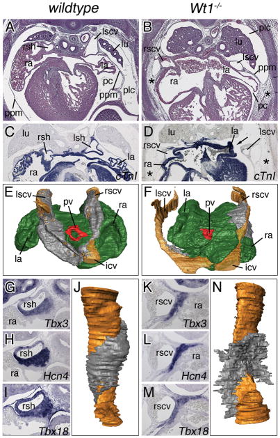
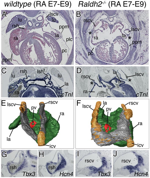
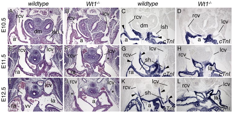
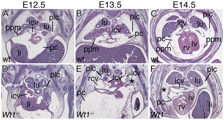
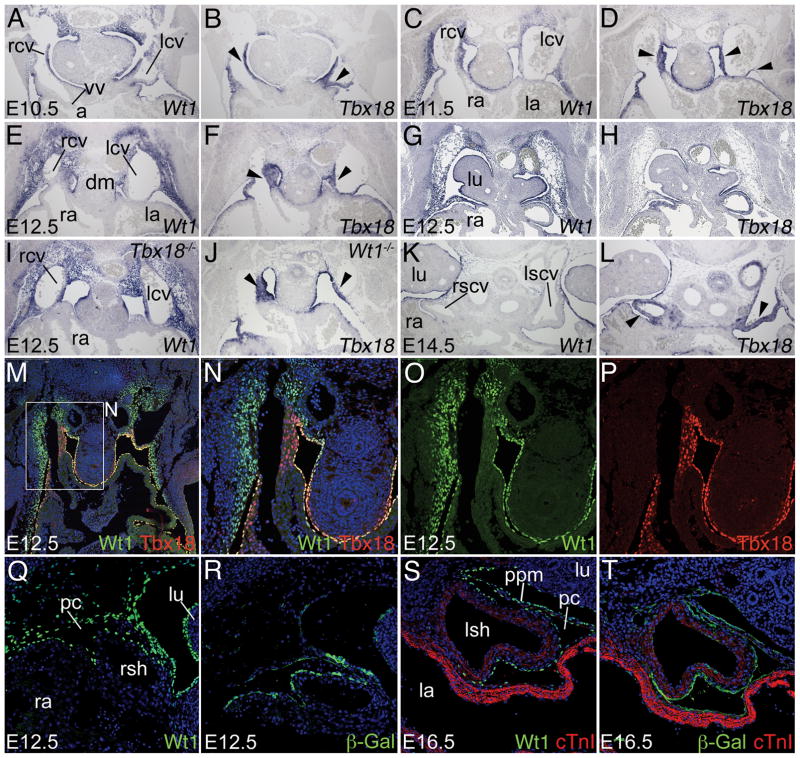
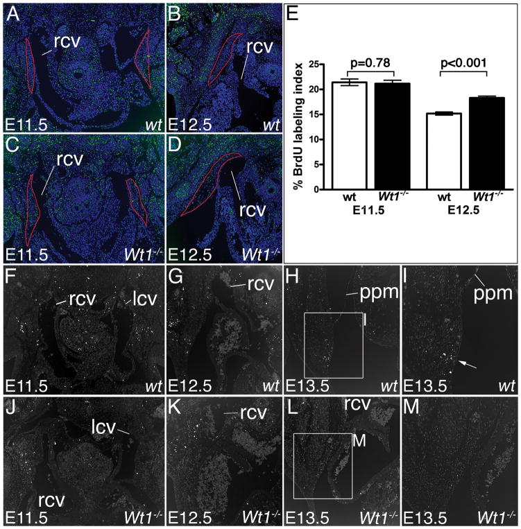
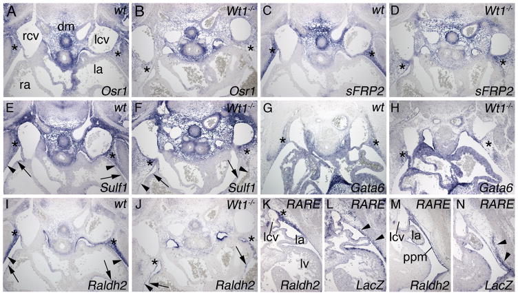
We analyzed embryos deficient for the Wt1 (Wilms tumor 1) gene and observed a failure to form myocardialized sinus horns. Instead, the cardinal veins become embedded laterally in the pleuropericardial membranes that remain tethered to the lateral body wall by the persisting subcoelomic mesenchyme, a finding that correlates with decreased apoptosis in this region. We show by expression analysis and lineage tracing studies that Wt1 is expressed in the subcoelomic mesenchyme surrounding the cardinal veins, but that this Wt1-positive mesenchyme does not contribute cells to the sinus horn myocardium. Expression of the Raldh2 (aldehyde dehydrogenase family 1, subfamily A2) gene was lost from this mesenchyme in Wt1(-/-) embryos. Phenotypic analysis of Raldh2 mutant mice rescued from early cardiac defects by retinoic acid food supply revealed defects of the venous pole and pericardium highly similar to those of Wt1(-/-) mice.
Pericardium and sinus horn formation are coupled and depend on the expansion and correct temporal release of pleuropericardial membranes from the underlying subcoelomic mesenchyme. Wt1 and downstream Raldh2/retinoic acid signaling are crucial regulators of this process. Thus, our results provide novel insight into the genetic and cellular pathways regulating the posterior extension of the mammalian heart and the formation of its coelomic lining.
心腔静脉极是先天性畸形和房性心律失常的常见焦点,但对于调节其发育的细胞和分子机制知之甚少。体循环静脉回流心肌(窦房结和窦角)仅在心发生晚期从心包间充质前体细胞池中形成。
分析指导胎儿窦角形成的细胞和分子机制。
我们分析了 Wt1(Wilms 肿瘤 1 基因)基因缺失的胚胎,观察到心肌化窦角形成失败。相反,心大静脉侧向嵌入胸心包膜中,胸心包膜通过持续的体腔下间充质仍然与侧体壁相连,这一发现与该区域细胞凋亡减少相关。我们通过表达分析和谱系追踪研究表明,Wt1 在围绕心大静脉的心包下间充质中表达,但该 Wt1 阳性间充质不会向窦角心肌提供细胞。Wt1(-/-) 胚胎中该间充质中的 Raldh2(醛脱氢酶家族 1,亚家族 A2)基因的表达丧失。用视黄酸食物供应从早期心脏缺陷中拯救出来的 Raldh2 突变小鼠的表型分析显示,静脉极和心包缺陷与 Wt1(-/-) 小鼠非常相似。
心包和窦角形成是偶联的,取决于胸心包膜从下面的体腔下间充质的扩张和正确的时间释放。Wt1 和下游的 Raldh2/视黄酸信号是该过程的关键调节因子。因此,我们的结果为调节哺乳动物心脏后部延伸和体腔衬里形成的遗传和细胞途径提供了新的见解。